


1
/
of
9
PayPal, credit cards. Download editable-PDF & invoice in 1 second!
GB 4789.14-2014 English PDF
GB 4789.14-2014 English PDF
Regular price
$115.00 USD
Regular price
Sale price
$115.00 USD
Unit price
/
per
Shipping calculated at checkout.
Couldn't load pickup availability
Delivery: 3 seconds. Download true-PDF + Invoice.
Get QUOTATION in 1-minute: Click GB 4789.14-2014
Historical versions: GB 4789.14-2014
Preview True-PDF (Reload/Scroll if blank)
GB 4789.14-2014: National Food Safety Standard -- Food Microbiological Examination -- Bacillus Cereus
GB 4789.14-2014
GB
NATIONAL STANDARD OF THE
PEOPLE’S REPUBLIC OF CHINA
National Food Safety Standard –
Food Microbiological Examination - Bacillus Cereus
ISSUED ON. DECEMBER 1, 2014
IMPLEMENTED ON. MAY 1, 2015
Issued by. National Health and Family Planning Commission of the
People’s Republic of China
Table of Contents
Foreword ... 3
1 Scope ... 4
2 Equipment and materials ... 4
3 Culture media and reagents ... 4
4 Plate count method of bacillus cereus (first method) ... 5
5 MPN count method of bacillus cereus (second method) ... 12
Annex A Culture media and reagents ... 15
Annex B Retrieval table of bacillus cereus most probable numbers ... 23
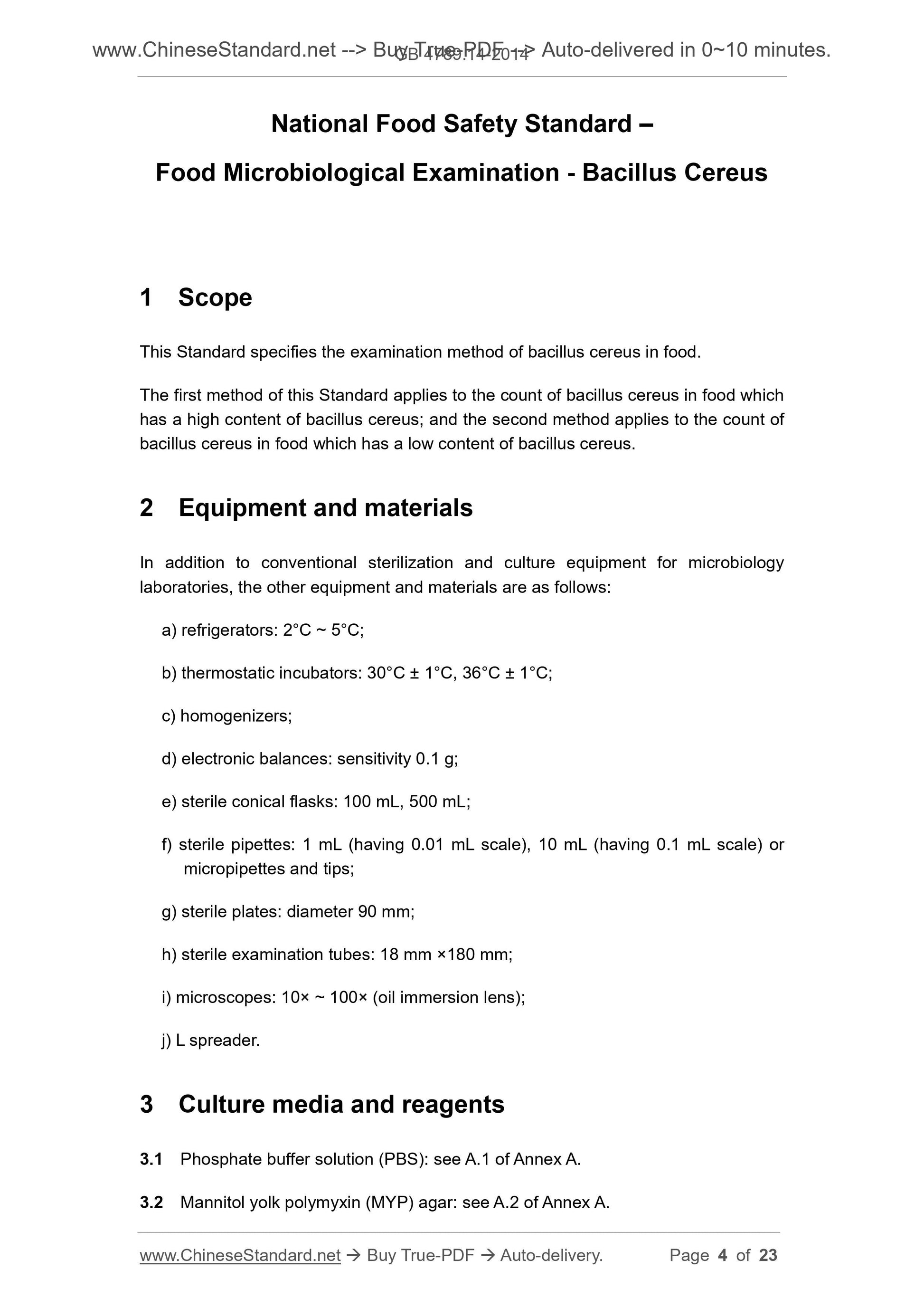
National Food Safety Standard –
Food Microbiological Examination - Bacillus Cereus
1 Scope
This Standard specifies the examination method of bacillus cereus in food.
The first method of this Standard applies to the count of bacillus cereus in food which
has a high content of bacillus cereus; and the second method applies to the count of
bacillus cereus in food which has a low content of bacillus cereus.
2 Equipment and materials
In addition to conventional sterilization and culture equipment for microbiology
laboratories, the other equipment and materials are as follows.
a) refrigerators. 2°C ~ 5°C;
b) thermostatic incubators. 30°C ± 1°C, 36°C ± 1°C;
c) homogenizers;
d) electronic balances. sensitivity 0.1 g;
e) sterile conical flasks. 100 mL, 500 mL;
f) sterile pipettes. 1 mL (having 0.01 mL scale), 10 mL (having 0.1 mL scale) or
micropipettes and tips;
g) sterile plates. diameter 90 mm;
h) sterile examination tubes. 18 mm ×180 mm;
i) microscopes. 10× ~ 100× (oil immersion lens);
j) L spreader.
3 Culture media and reagents
3.1 Phosphate buffer solution (PBS). see A.1 of Annex A.
3.2 Mannitol yolk polymyxin (MYP) agar. see A.2 of Annex A.
4.2.3 Sample dilution
Absorb 1 mL of the 1.10 sample homogeneous solution of 4.2.2 to add to a dilution
tube containing 9 mL of PBS or normal saline; fully mix up to make 1.100 sample
homogeneous solution. In accordance with the estimation of sample contamination
degree, operate as above and make into ten-fold incremental serial dilution sample
homogeneous solution in succession. After each dilution, replace one 1 mL sterile
pipette or tip.
4.2.4 Sample inoculation
In accordance with the estimation of sample contamination degree, select 2 ~ 3 sample
homogeneous solutions of appropriate dilution (liquid samples can include primary
liquid); transfer the inoculum sizes of 0.3 mL, 0.3 mL and 0.4 mL to three MYP agar
plates respectively; then use a sterile L spreader to apply them to the whole plates;
and pay attention to not touching the edge of the plates. Before use, if there are water
drops on the surface of the MYP agar plates, they can be placed in an incubator to dry
at 25°C ~ 50°C until the water drops on the surface of the plates disappear.
4.2.5 Isolation and culture
4.2.5.1 Isolation
Under normal conditions, allow plates to stand for 10 min after applying. If sample
solution is difficult to absorb, it can be placed in an incubator to culture for 1 h at 30°C
± 1°C; turn over plates after sample homogeneous solution is evenly absorbed; place
them upside down in the incubator; culture for 24 h ± 2 h at 30°C ± 1°C. If the colonies
are not typical, continue culturing for 24 h ± 2 h before observing. On MYP agar plates,
typical colonies are of a faint pink colour (indicating unfermented mannitol) and there
are white to pale orchid pink precipitation ring (indicating the production of lecithinase).
4.2.5.2 Pure culture
Select at least 5 typical colonies (select all if less than 5) from each plate (complying
with the requirements of 4.4.1.1); inoculate them by streaking in nutrient agar plates
for pure culture; culture for 24 h ± 2 h at 30°C ± 1°C to carry out confirmatory test. On
nutrient agar plates, typical colonies are offwhite, occasionally yellowish green, non-
transparent, ground glass shaped or melting wax shaped in surface roughness, usually
flared on the edge and of diameter 4 mm ~ 10 mm.
4.3 Confirmatory appraisal
4.3.1 Dyeing microscopic examination
Pick single colonies of pure culture for Gram’s microscopic examination. Bacillus
cereus is Gram positive bacillus of size (1 μm ~ 1.3 μm) ×(3 μm ~ 5 μm); the spore is
oval which is located at the centre or one end of thallus, not expanding on thallus; both
ends of thallus are flat, normally arranged in the shape of short chains or long chains.
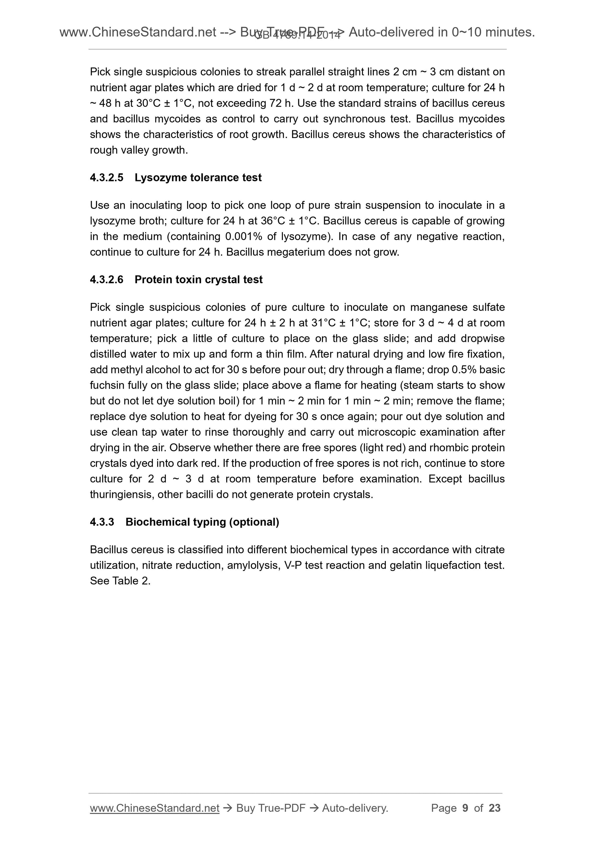
Pick single suspicious colonies to streak parallel straight lines 2 cm ~ 3 cm distant on
nutrient agar plates which are dried for 1 d ~ 2 d at room temperature; culture for 24 h
~ 48 h at 30°C ± 1°C, not exceeding 72 h. Use the standard strains of bacillus cereus
and bacillus mycoides as control to carry out synchronous test. Bacillus mycoides
shows the characteristics of root growth. Bacillus cereus shows the characteristics of
rough valley growth.
4.3.2.5 Lysozyme tolerance test
Use an inoculating loop to pick one loop of pure strain suspension to inoculate in a
lysozyme broth; culture for 24 h at 36°C ± 1°C. Bacillus cereus is capable of growing
in the medium (containing 0.001% of lysozyme). In case of any negative reaction,
continue to culture for 24 h. Bacillus megaterium does not grow.
4.3.2.6 Protein toxin crystal test
Pick single suspicious colonies of pure culture to inoculate on manganese sulfate
nutrient agar plates; culture for 24 h ± 2 h at 31°C ± 1°C; store for 3 d ~ 4 d at room
temperature; pick a little of culture to place on the glass slide; and add dropwise
distilled water to mix up and form a thin film. After natural drying and low fire fixation,
add methyl alcohol to act for 30 s before pour out; dry through a flame; drop 0.5% basic
fuchsin fully on the glass slide; place above a flame for heating (steam starts to show
but do not let dye solution boil) for 1 min ~ 2 min for 1 min ~ 2 min; remove the flame;
replace dye solution to heat for dyeing for 30 s once again; pour out dye solution and
use clean tap water to rinse thoroughly and carry out microscopic examination after
drying in the air. Observe whether there are free spores (light red) and rhombic protein
crystals dyed into dark red. If the production of free spores is not rich, continue to store
culture for 2 d ~ 3 d at room temperature before examination. Except bacillus
thuringiensis, other bacilli do not generate protein crystals.
4.3.3 Biochemical typing (optional)
Bacillus cereus is classified into different biochemical types in accordance with citrate
utilization, nitrate reduction, amylolysis, V-P test reaction and gelatin liquefaction test.
See Table 2.
Annex A
Culture media and reagents
A.1 Phosphate buffer solution (PBS)
A.1.1 Composition
Potassium dihydrogen phosphate 34.0 g
Distilled water 500.0 mL
A.1.2 Preparation
Stock solution. weigh 34.0 g of potassium dihydrogen phosphate to dissolve in 500 mL
of distilled water; use about 175 mL of 1 mol/L sodium hydroxide solution to adjust pH
to 7.2; use distilled water to dilute to 1 000 mL before storing in a refrigerator.
Dilute solution. take 1.25 mL of stock solution; use distilled water to dilute to 1 000 mL;
load in appropriate containers; carry out autoclaved sterilization for 15 min at 121°C.
A.2 Mannitol yolk polymyxin (MYP) agar
A.2.1 Composition
Peptone 10.0 g
Beef powder 1.0 g
D-mannitol 10.0 g
Sodium chlori...
Get QUOTATION in 1-minute: Click GB 4789.14-2014
Historical versions: GB 4789.14-2014
Preview True-PDF (Reload/Scroll if blank)
GB 4789.14-2014: National Food Safety Standard -- Food Microbiological Examination -- Bacillus Cereus
GB 4789.14-2014
GB
NATIONAL STANDARD OF THE
PEOPLE’S REPUBLIC OF CHINA
National Food Safety Standard –
Food Microbiological Examination - Bacillus Cereus
ISSUED ON. DECEMBER 1, 2014
IMPLEMENTED ON. MAY 1, 2015
Issued by. National Health and Family Planning Commission of the
People’s Republic of China
Table of Contents
Foreword ... 3
1 Scope ... 4
2 Equipment and materials ... 4
3 Culture media and reagents ... 4
4 Plate count method of bacillus cereus (first method) ... 5
5 MPN count method of bacillus cereus (second method) ... 12
Annex A Culture media and reagents ... 15
Annex B Retrieval table of bacillus cereus most probable numbers ... 23
National Food Safety Standard –
Food Microbiological Examination - Bacillus Cereus
1 Scope
This Standard specifies the examination method of bacillus cereus in food.
The first method of this Standard applies to the count of bacillus cereus in food which
has a high content of bacillus cereus; and the second method applies to the count of
bacillus cereus in food which has a low content of bacillus cereus.
2 Equipment and materials
In addition to conventional sterilization and culture equipment for microbiology
laboratories, the other equipment and materials are as follows.
a) refrigerators. 2°C ~ 5°C;
b) thermostatic incubators. 30°C ± 1°C, 36°C ± 1°C;
c) homogenizers;
d) electronic balances. sensitivity 0.1 g;
e) sterile conical flasks. 100 mL, 500 mL;
f) sterile pipettes. 1 mL (having 0.01 mL scale), 10 mL (having 0.1 mL scale) or
micropipettes and tips;
g) sterile plates. diameter 90 mm;
h) sterile examination tubes. 18 mm ×180 mm;
i) microscopes. 10× ~ 100× (oil immersion lens);
j) L spreader.
3 Culture media and reagents
3.1 Phosphate buffer solution (PBS). see A.1 of Annex A.
3.2 Mannitol yolk polymyxin (MYP) agar. see A.2 of Annex A.
4.2.3 Sample dilution
Absorb 1 mL of the 1.10 sample homogeneous solution of 4.2.2 to add to a dilution
tube containing 9 mL of PBS or normal saline; fully mix up to make 1.100 sample
homogeneous solution. In accordance with the estimation of sample contamination
degree, operate as above and make into ten-fold incremental serial dilution sample
homogeneous solution in succession. After each dilution, replace one 1 mL sterile
pipette or tip.
4.2.4 Sample inoculation
In accordance with the estimation of sample contamination degree, select 2 ~ 3 sample
homogeneous solutions of appropriate dilution (liquid samples can include primary
liquid); transfer the inoculum sizes of 0.3 mL, 0.3 mL and 0.4 mL to three MYP agar
plates respectively; then use a sterile L spreader to apply them to the whole plates;
and pay attention to not touching the edge of the plates. Before use, if there are water
drops on the surface of the MYP agar plates, they can be placed in an incubator to dry
at 25°C ~ 50°C until the water drops on the surface of the plates disappear.
4.2.5 Isolation and culture
4.2.5.1 Isolation
Under normal conditions, allow plates to stand for 10 min after applying. If sample
solution is difficult to absorb, it can be placed in an incubator to culture for 1 h at 30°C
± 1°C; turn over plates after sample homogeneous solution is evenly absorbed; place
them upside down in the incubator; culture for 24 h ± 2 h at 30°C ± 1°C. If the colonies
are not typical, continue culturing for 24 h ± 2 h before observing. On MYP agar plates,
typical colonies are of a faint pink colour (indicating unfermented mannitol) and there
are white to pale orchid pink precipitation ring (indicating the production of lecithinase).
4.2.5.2 Pure culture
Select at least 5 typical colonies (select all if less than 5) from each plate (complying
with the requirements of 4.4.1.1); inoculate them by streaking in nutrient agar plates
for pure culture; culture for 24 h ± 2 h at 30°C ± 1°C to carry out confirmatory test. On
nutrient agar plates, typical colonies are offwhite, occasionally yellowish green, non-
transparent, ground glass shaped or melting wax shaped in surface roughness, usually
flared on the edge and of diameter 4 mm ~ 10 mm.
4.3 Confirmatory appraisal
4.3.1 Dyeing microscopic examination
Pick single colonies of pure culture for Gram’s microscopic examination. Bacillus
cereus is Gram positive bacillus of size (1 μm ~ 1.3 μm) ×(3 μm ~ 5 μm); the spore is
oval which is located at the centre or one end of thallus, not expanding on thallus; both
ends of thallus are flat, normally arranged in the shape of short chains or long chains.
Pick single suspicious colonies to streak parallel straight lines 2 cm ~ 3 cm distant on
nutrient agar plates which are dried for 1 d ~ 2 d at room temperature; culture for 24 h
~ 48 h at 30°C ± 1°C, not exceeding 72 h. Use the standard strains of bacillus cereus
and bacillus mycoides as control to carry out synchronous test. Bacillus mycoides
shows the characteristics of root growth. Bacillus cereus shows the characteristics of
rough valley growth.
4.3.2.5 Lysozyme tolerance test
Use an inoculating loop to pick one loop of pure strain suspension to inoculate in a
lysozyme broth; culture for 24 h at 36°C ± 1°C. Bacillus cereus is capable of growing
in the medium (containing 0.001% of lysozyme). In case of any negative reaction,
continue to culture for 24 h. Bacillus megaterium does not grow.
4.3.2.6 Protein toxin crystal test
Pick single suspicious colonies of pure culture to inoculate on manganese sulfate
nutrient agar plates; culture for 24 h ± 2 h at 31°C ± 1°C; store for 3 d ~ 4 d at room
temperature; pick a little of culture to place on the glass slide; and add dropwise
distilled water to mix up and form a thin film. After natural drying and low fire fixation,
add methyl alcohol to act for 30 s before pour out; dry through a flame; drop 0.5% basic
fuchsin fully on the glass slide; place above a flame for heating (steam starts to show
but do not let dye solution boil) for 1 min ~ 2 min for 1 min ~ 2 min; remove the flame;
replace dye solution to heat for dyeing for 30 s once again; pour out dye solution and
use clean tap water to rinse thoroughly and carry out microscopic examination after
drying in the air. Observe whether there are free spores (light red) and rhombic protein
crystals dyed into dark red. If the production of free spores is not rich, continue to store
culture for 2 d ~ 3 d at room temperature before examination. Except bacillus
thuringiensis, other bacilli do not generate protein crystals.
4.3.3 Biochemical typing (optional)
Bacillus cereus is classified into different biochemical types in accordance with citrate
utilization, nitrate reduction, amylolysis, V-P test reaction and gelatin liquefaction test.
See Table 2.
Annex A
Culture media and reagents
A.1 Phosphate buffer solution (PBS)
A.1.1 Composition
Potassium dihydrogen phosphate 34.0 g
Distilled water 500.0 mL
A.1.2 Preparation
Stock solution. weigh 34.0 g of potassium dihydrogen phosphate to dissolve in 500 mL
of distilled water; use about 175 mL of 1 mol/L sodium hydroxide solution to adjust pH
to 7.2; use distilled water to dilute to 1 000 mL before storing in a refrigerator.
Dilute solution. take 1.25 mL of stock solution; use distilled water to dilute to 1 000 mL;
load in appropriate containers; carry out autoclaved sterilization for 15 min at 121°C.
A.2 Mannitol yolk polymyxin (MYP) agar
A.2.1 Composition
Peptone 10.0 g
Beef powder 1.0 g
D-mannitol 10.0 g
Sodium chlori...
Share