1
/
su
6
PayPal, credit cards. Download editable-PDF and invoice in 1 second!
YY/T 0767-2023 English PDF (YYT0767-2023)
YY/T 0767-2023 English PDF (YYT0767-2023)
Prezzo di listino
$230.00 USD
Prezzo di listino
Prezzo scontato
$230.00 USD
Prezzo unitario
/
per
Spese di spedizione calcolate al check-out.
Impossibile caricare la disponibilità di ritiro
Delivery: 3 seconds. Download true-PDF + Invoice.
Get QUOTATION in 1-minute: Click YY/T 0767-2023
Historical versions: YY/T 0767-2023
Preview True-PDF (Reload/Scroll if blank)
YY/T 0767-2023: General technical requirements for color ultrasound imaging equipment
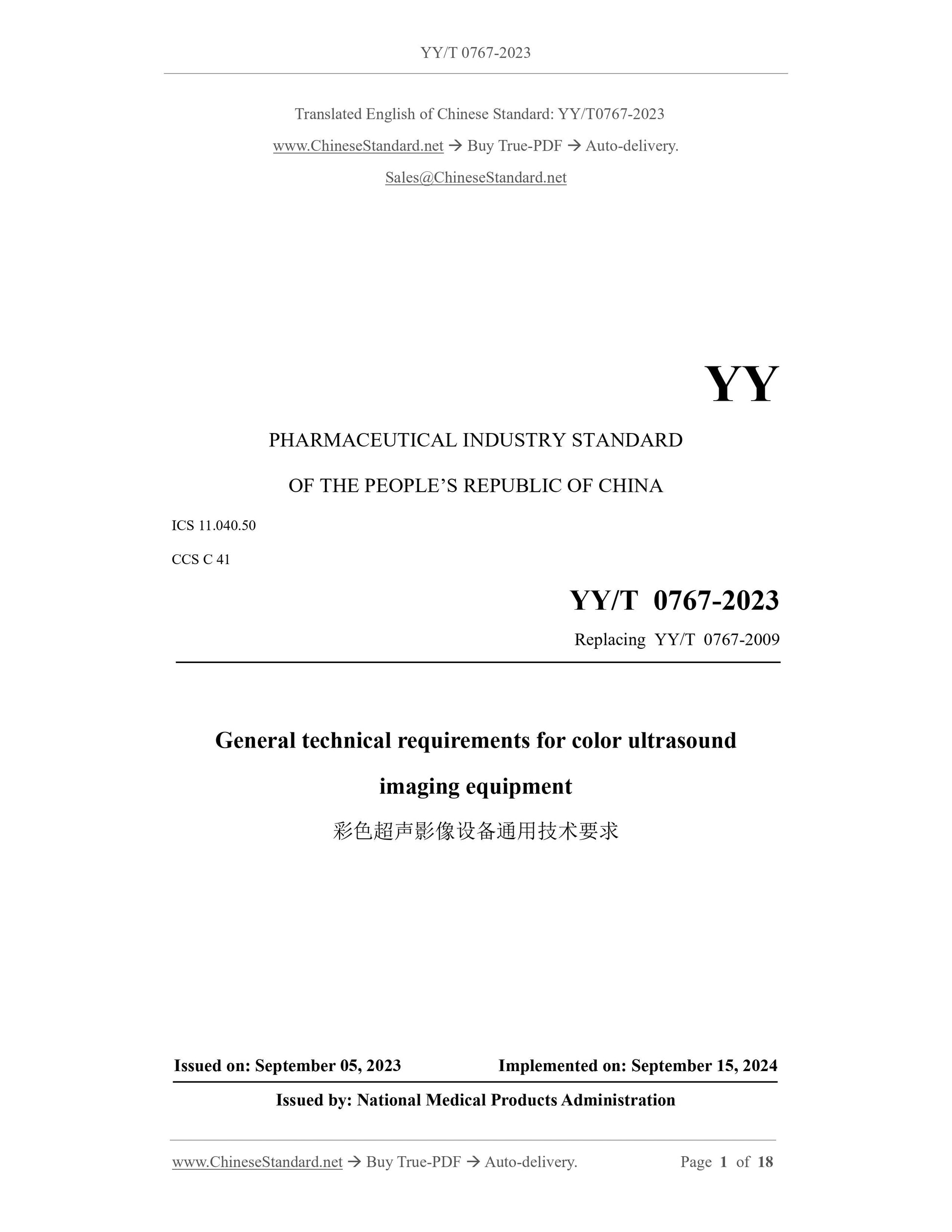
YY/T 0767-2023
YY
PHARMACEUTICAL INDUSTRY STANDARD
OF THE PEOPLE’S REPUBLIC OF CHINA
ICS 11.040.50
CCS C 41
Replacing YY/T 0767-2009
General technical requirements for color ultrasound
imaging equipment
ISSUED ON: SEPTEMBER 05, 2023
IMPLEMENTED ON: SEPTEMBER 15, 2024
Issued by: National Medical Products Administration
Table of Contents
Foreword ... 3
1 Scope ... 5
2 Normative references ... 5
3 Terms and definitions ... 6
4 Requirements ... 7
5 Test methods ... 8
Annex A (informative) Doppler performance parameters and test equipment ... 13
Annex B (informative) Considerations for setting various parameters during color
ultrasound testing ... 17
General technical requirements for color ultrasound
imaging equipment
1 Scope
This document specifies the requirements for color ultrasound imaging equipment
(referred to as "color ultrasound") and describes the corresponding test methods.
This document applies to color ultrasound imaging equipment.
2 Normative references
The following referenced documents are indispensable for the application of this
document. For dated references, only the edition cited applies. For undated references,
the latest edition of the referenced document (including any amendments) applies.
GB 9706.1, Medical electrical equipment -- Part 1: General requirements for basic
safety and essential performance
GB 9706.237, Medical electrical equipment -- Part 2-37: Particular requirements
for the basic safety and essential performance of ultrasonic medical diagnostic and
monitoring equipment
GB 10152, B mode ultrasonic diagnostic equipment
GB/T 14710, Environmental requirement and test methods for medical electrical
equipment
YY 9706.102, Medical electrical equipment -- Part 1-2: General requirements for
basic safety and essential performance -- Collateral standard: Electromagnetic
compatibility -- Requirements and tests
YY/T 0458, Technical requirements for ultrasonically blood-mimicking Doppler
phantom
YY/T 0705, Ultrasonics. Continuous-wave Doppler systems. Test procedures
YY/T 1420, Environmental requirement and test methods for medical ultrasonic
equipment
blood flow model or the string Doppler test piece and the set speed.
3.7 directional discrimination
The ability that color Doppler ultrasound identifies the direction of blood flow and
expresses the direction in terms of the color of the blood flow map and/or the position
of the Doppler spectrum relative to the baseline.
4 Requirements
4.1 General
The manufacturer shall publish the indicators of all applicable technical parameters in
this chapter in the accompanying documents according to the actual probe configuration
of the equipment.
For multi-purpose color ultrasound, manufacturers can give different indicators for the
same probe for different expected application ranges.
4.2 Color Doppler mode
4.2.1 The penetration depth of Doppler of color ultrasound shall comply with the
specifications published by the manufacturer in the accompanying document.
4.2.2 The color Doppler image shall match the grayscale image of the pipe in which it
is located.
4.2.3 It shall have directional discrimination.
4.3 Spectral Doppler mode
4.3.1 The penetration depth of Doppler of color ultrasound shall comply with the
specifications published by the manufacturer in the accompanying document.
4.3.2 The error of flow velocity shall be within ±20%.
4.3.3 The cursor position of the sampling area in pulse wave Doppler mode shall be
accurate.
4.4 Power Doppler mode
4.4.1 The penetration depth of power Doppler of color ultrasound shall comply with the
specifications published by the manufacturer in the accompanying document.
4.4.2 The power Doppler image shall match the grayscale image of the pipe in which it
is located.
4.5 Safety
Color Doppler ultrasound is generally equipped with multiple probes. A probe may have
several intended uses. There are many combinations of color ultrasound host control
terminal (acoustic output, gain, focus position, etc.) settings and probe selection. It is
not possible to test all combinations of states. In this document, the main unit and probe
combinations are tested only in the specified settings.
When testing, it is recommended to simulate the most commonly used states of the
device and probe in expected clinical imaging.
During the test, it is recommended that the manufacturer provide the best setting status
in the accompanying document, or use the one-click optimization setting function of
color ultrasound, and avoid setting the device's display brightness, contrast, etc. to the
extreme state, see Annex B.
5.2 Color Doppler mode
5.2.1 The probe is well coupled to the simulated blood fluid model acoustic window.
The ultrasound beam axis is aligned with the simulated blood vessels in the phantom.
Under the prescribed settings, keep the simulated blood vessel image clearly visible and
obtain clear color Doppler images. Move the probe in a deeper direction along the
direction of the simulated blood vessel until the colored signal disappears. Thereafter
the probe is moved back to the position before the color signal disappeared. Freeze the
image. Use the ranging function of the equipment to measure the distance between the
acoustic window and the farthest end of the inner wall at this time, which is the
penetration depth of color Doppler of color ultrasound.
5.2.2 The probe is well coupled to the simulated blood fluid model acoustic window.
The ultrasound beam axis is aligned with the simulated blood vessels in the phantom.
Under the prescribed settings, keep the simulated blood vessel image clearly visible and
obtain clear color Doppler images. Observe and verify that the color blood flow image
shall be consistent with the grayscale image of the pipeline where it is located, that is,
there is no obvious lack of filling, no obvious misalignment, and no obvious color
overflow.
5.2.3 The probe is well coupled to the simulated blood fluid model acoustic window.
The ultrasound beam axis is aligned with the simulated blood vessels in the phantom.
Under the prescribed settings, keep the simulated blood vessel image clearly visible and
obtain clear color Doppler images. By changing the blood flow direction relative to the
probe, the device shall be able to correctly identify the blood flow direction without
aliasing.
For a blood-simulating fluid model containing two parallel pipes with opposite
directions, the beam scanning plane can be used to intercept the cross-sections of the
two pipes for inspection.
Convex array and phased array probes can also use simulated blood fluid models
containing horizontal pipes. It is not necessary to turn the probe at this time. Hold the
5.3.3 The probe is well coupled to the simulated blood fluid model acoustic window.
The ultrasound beam axis is aligned with the simulated blood vessels in the phantom.
Under the prescribed settings, keep the simulated blood vessel image clearly visible and
obtain a clear spectral Doppler signal. First, place the sampling area outside the
superficial wall of the simulated blood vessel. The Doppler spectrum shall not be
displayed at this time. Then, in small steps of 0.5 mm or 1 mm, the sampling area is
advanced from shallow to deep, across the blood vessel wall toward the blood vessel
center, and finally to the outside of the deep blood vessel wall. The Doppler spectrum
display shall be from weak to ...
Share
