1
/
の
12
PayPal, credit cards. Download editable-PDF & invoice in 1 second!
YY 0831.1-2011 English PDF (YY0831.1-2011)
YY 0831.1-2011 English PDF (YY0831.1-2011)
通常価格
$150.00 USD
通常価格
セール価格
$150.00 USD
単価
/
あたり
配送料はチェックアウト時に計算されます。
受取状況を読み込めませんでした
Delivery: 3 seconds. Download true-PDF + Invoice.
Get QUOTATION in 1-minute: Click YY 0831.1-2011
Historical versions: YY 0831.1-2011
Preview True-PDF (Reload/Scroll if blank)
YY 0831.1-2011: Stereotactic radiotherapy system with gamma beam - Part 1: Multi-source stereotactic radiotherapy system with gamma beam for head lesion
YY 0831‐2011
YY
ICS 11.040.50
C 43
PHARMACEUTICAL INDUSTRY STANDARD
OF THE PEOPLE’S REPUBLIC OF CHINA
Stereotactic radiotherapy system with gamma
beam — Part 1. Multi-source stereotactic
radiotherapy system with gamma beam for head
lesion
ISSUED ON. DECEMBER 31, 2011
IMPLEMENTED ON. JUNE 1, 2013
Issued by. State Food and Drug Administration
Table of Contents
Foreword ... 3
1 Scope ... 4
2 Normative references ... 4
3 Terms and definitions ... 5
4 Requirements ... 5
5 Test methods ... 8
Annex A (Informative) Materials, structure and size of special spherical die
body and special focus measuring tools for test ... 15
Foreword
All technical contents of this Part are mandatory.
YY 0831 is consisted of 2 parts as follows.
— Part 1. Multi-source stereotactic radiotherapy system with gamma beam for head
lesion;
— Part 2. Multi-source stereotactic radiotherapy system with gamma beam for body
lesion.
This Part is part 1 of YY 0831.
This Part is drafted according to the rules specified in GB/T 1.1-2009.
Please note that some contents in this Document may involve patents. The issuing
authority of this Document does not undertake the responsibilities to identify these
patents.
The part was proposed by China Food and Drug Administration.
The part shall be under the jurisdiction of National Standardization Technical Committee of
Medical Appliances Radiotherapy and Technical Committee of Nuclear Medicine and
Radiation Dosimetry Equipment (SAC/TC10/SC3).
Drafting organization of this Part. Beijing Institute of Medical Device Testing, Shenzhen
Haibo Technology Co., Ltd., Medical Science and Technology Development (Shenzhen) Co.,
Ltd. (MASEP), Shenzhen Shengai Medical Technology Development Co., Ltd., and
Shenzhen Zunrui Technology Co., Ltd.
The main drafters of the part. Zhang Zhaoyuan, Zhang Xin, Song Lianyou, Chen Jing, Zhu
Weiqun, Xu Tao, Zheng Tie, and Liu Guangwu.
Stereotactic radiotherapy system with gamma beam —
Part 1. Multi-source stereotactic radiotherapy system
with gamma beam for head lesion
1 Scope
This Part of YY 0831 specifies the scope, terms, definitions, requirements, and test
methods of the multi-source stereotactic radiotherapy system with gamma beam for head
lesion.
This Part applies to the multi-source stereotactic radiotherapy system with gamma beam
for head lesion (hereinafter referred to as the system). The system simultaneously uses
multiple 60Co sealed radioactive sources (it may be dynamic or static) to conduct beaming
irradiation to the head lesion.
2 Normative references
The articles contained in the following documents have become part of this Document
when they are quoted herein. For the dated documents so quoted, all the modifications
(Including all corrections) or revisions made thereafter shall be applicable to this
Document.
GB 9706.1-2007 Medical electrical equipment - Part 1. General requirements for safety
GB 9706.15-2008 Medical electrical equipment - Part 1. General requirements for safety
- Collateral standard. Safety requirements for medical electrical systems
GB 9706.17-2009 Medical electrical equipment - Part 2. Particular requirements for the
safety of gamma beam therapy equipment
GB/T 17857-1999 Medical radiology – Terminology (Equipment for radiotherapy, nuclear
medicine and radiation dosimetry)
GB/T 18987-2003 Radiotherapy equipment - Coordinates, movements and scales
YY 0637-2012 Medical electrical equipment - Requirements for the safety of
radiotherapy therapeutic planning systems
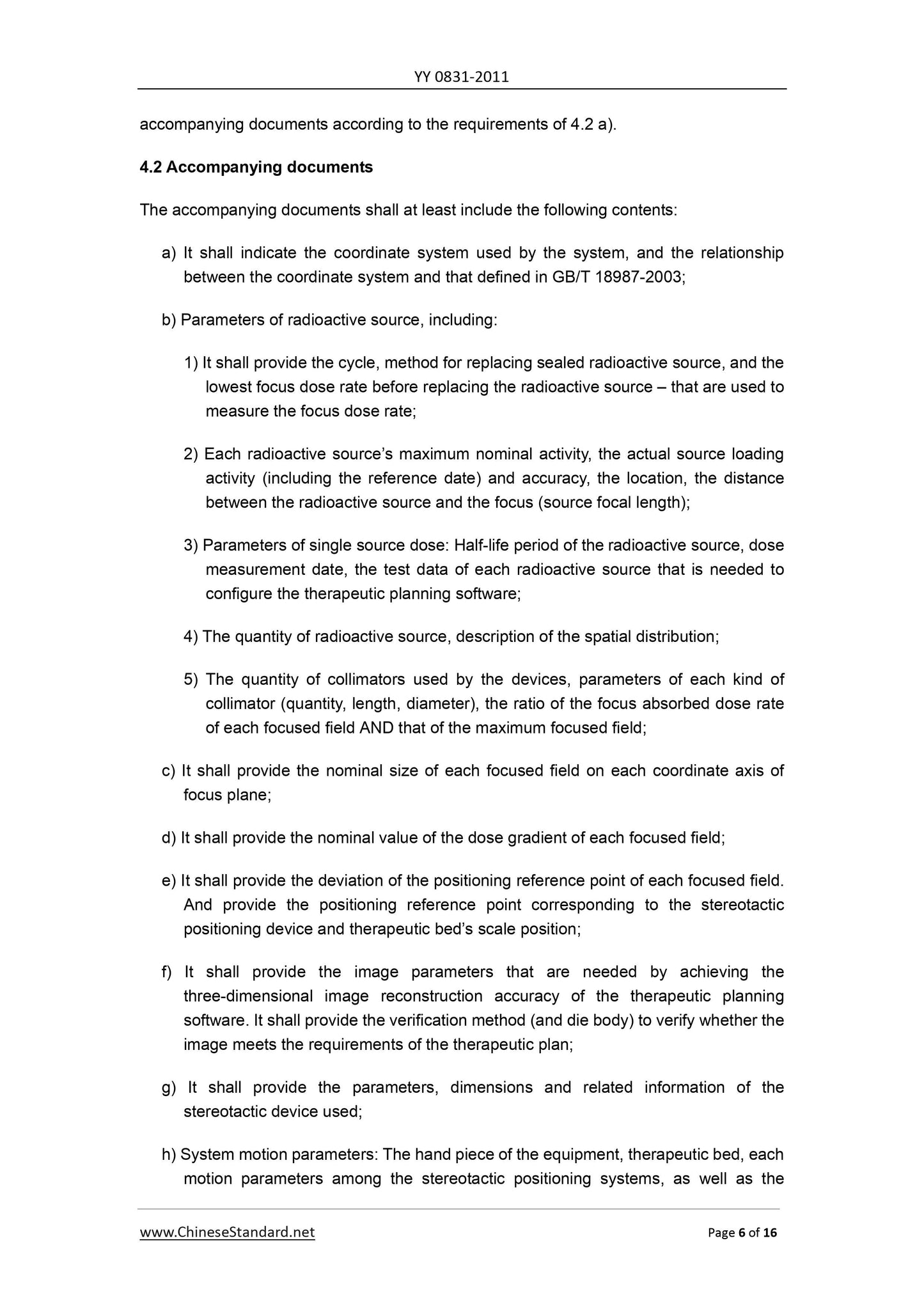
c) For the focused field of which the nominal size is greater than 20 mm and less than
or equal to 30 mm, the dose gradient of each focused field shall not exceed 10 mm.
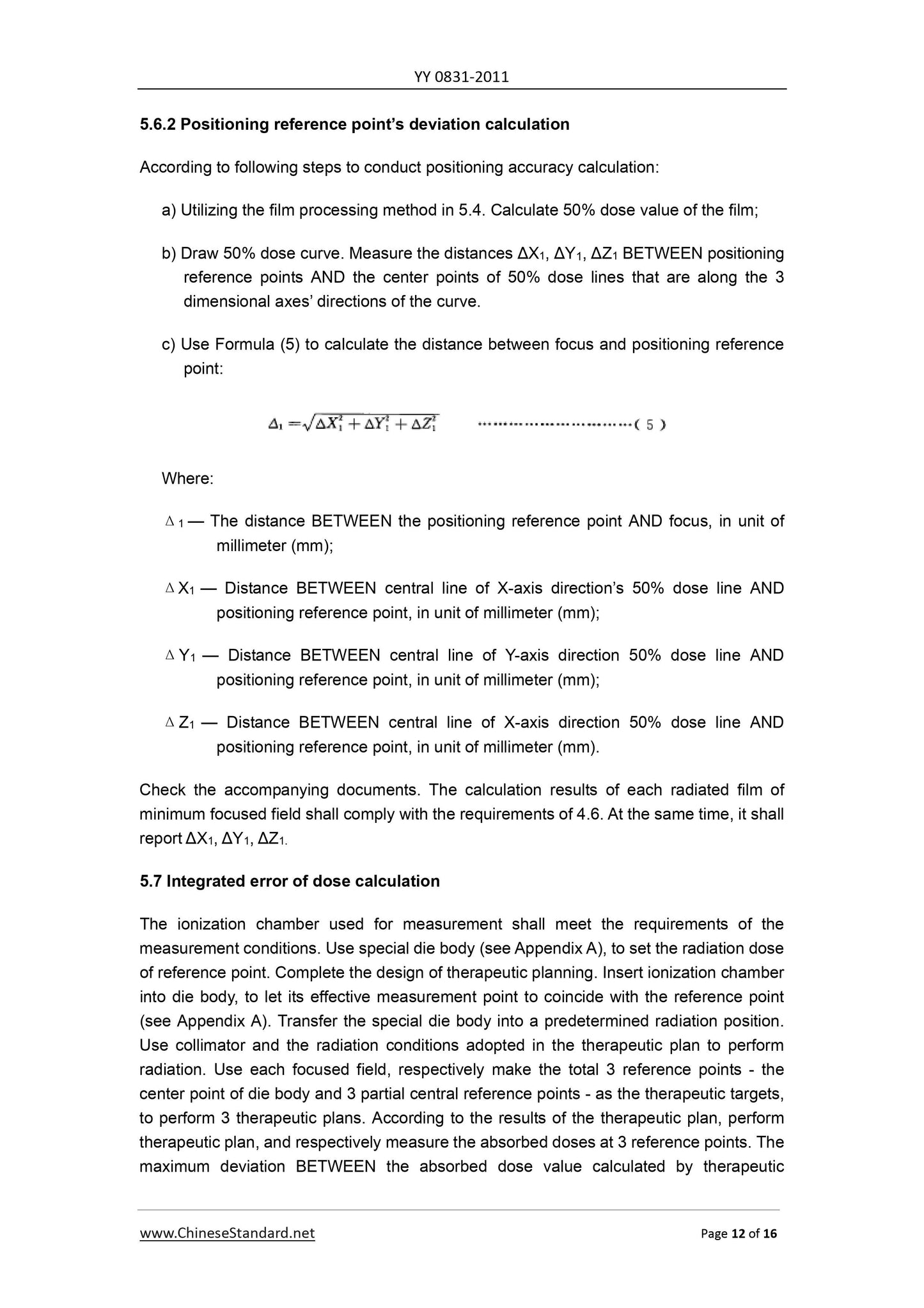
4.6 Deviation of positioning reference point
Deviation of the positioning reference point of the system shall meet the following
requirements.
a) For the minimum focused field, the deviation between the positioning reference point
and the focus shall be less than or equal to 0.5 mm;
b) The deviations of positioning reference point for each focused field shall be included
in accompanying documents.
4.7 Integrated error of dose calculation
For each focused field, the error between the absorbent dose value calculated by the
therapeutic planning software AND the actual measured value shall not exceed ±5%.
4.8 Position error of therapeutic plan reference point
Reconstruction position error of therapeutic planning software shall not exceed 1.5 mm.
4.9 Safety requirements
The system shall meet the requirements of GB 9706.1-2007, GB 9706.15-2008, GB
9706.17-2009 and YY 0637-2012.
5 Test methods
5.1 Coordinate system
Check the accompanying documents. It shall comply with the requirements of 4.1.
5.2 Accompanying documents
Check the accompanying documents. It shall comply with the requirements of 4.2.
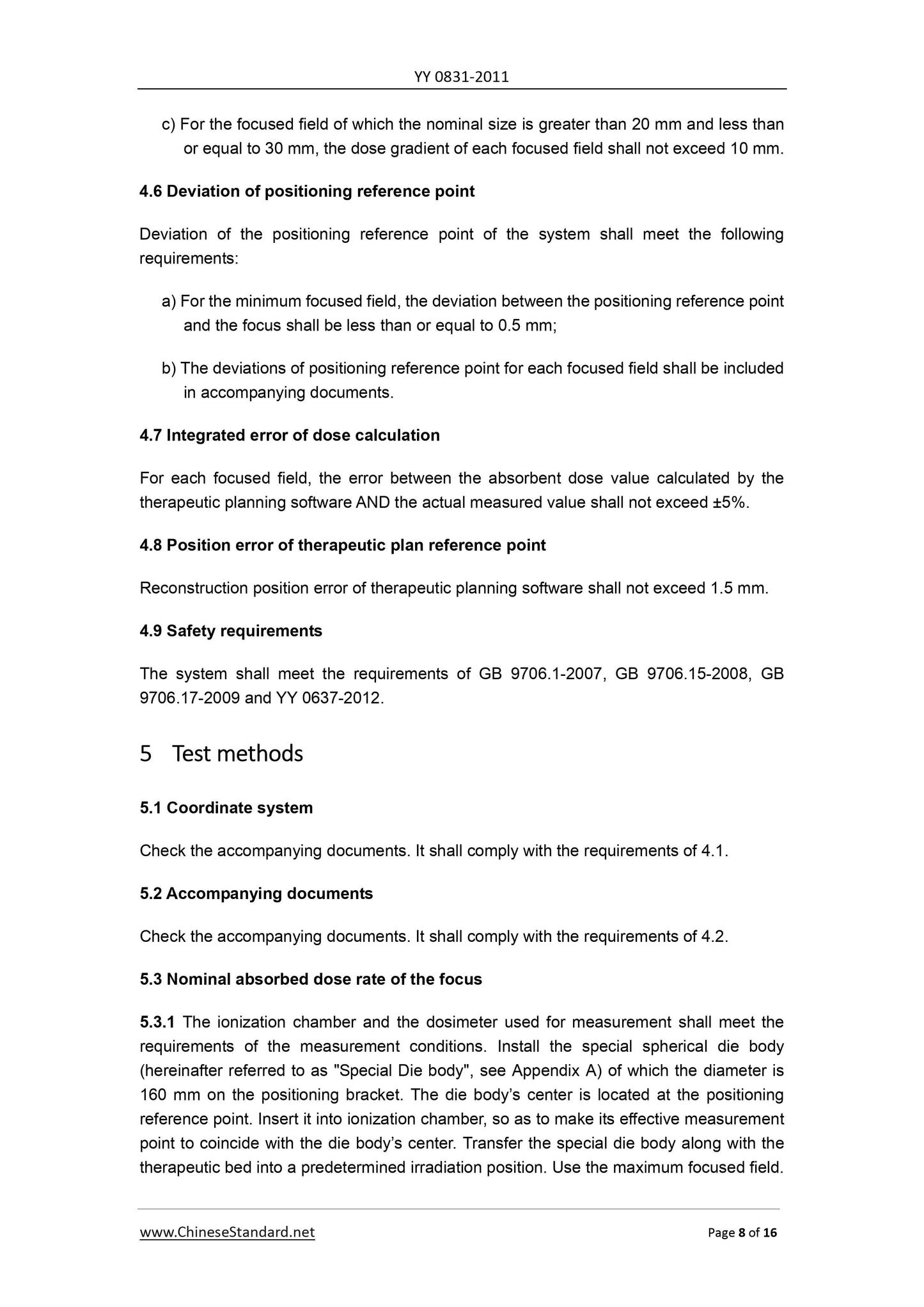
5.3 Nominal absorbed dose rate of the focus
5.3.1 The ionization chamber and the dosimeter used for measurement shall meet the
requirements of the measurement conditions. Install the special spherical die body
(hereinafter referred to as "Special Die body", see Appendix A) of which the diameter is
160 mm on the positioning bracket. The die body’s center is located at the positioning
reference point. Insert it into ionization chamber, so as to make its effective measurement
point to coincide with the die body’s center. Transfer the special die body along with the
therapeutic bed into a predetermined irradiation position. Use the maximum focused field.
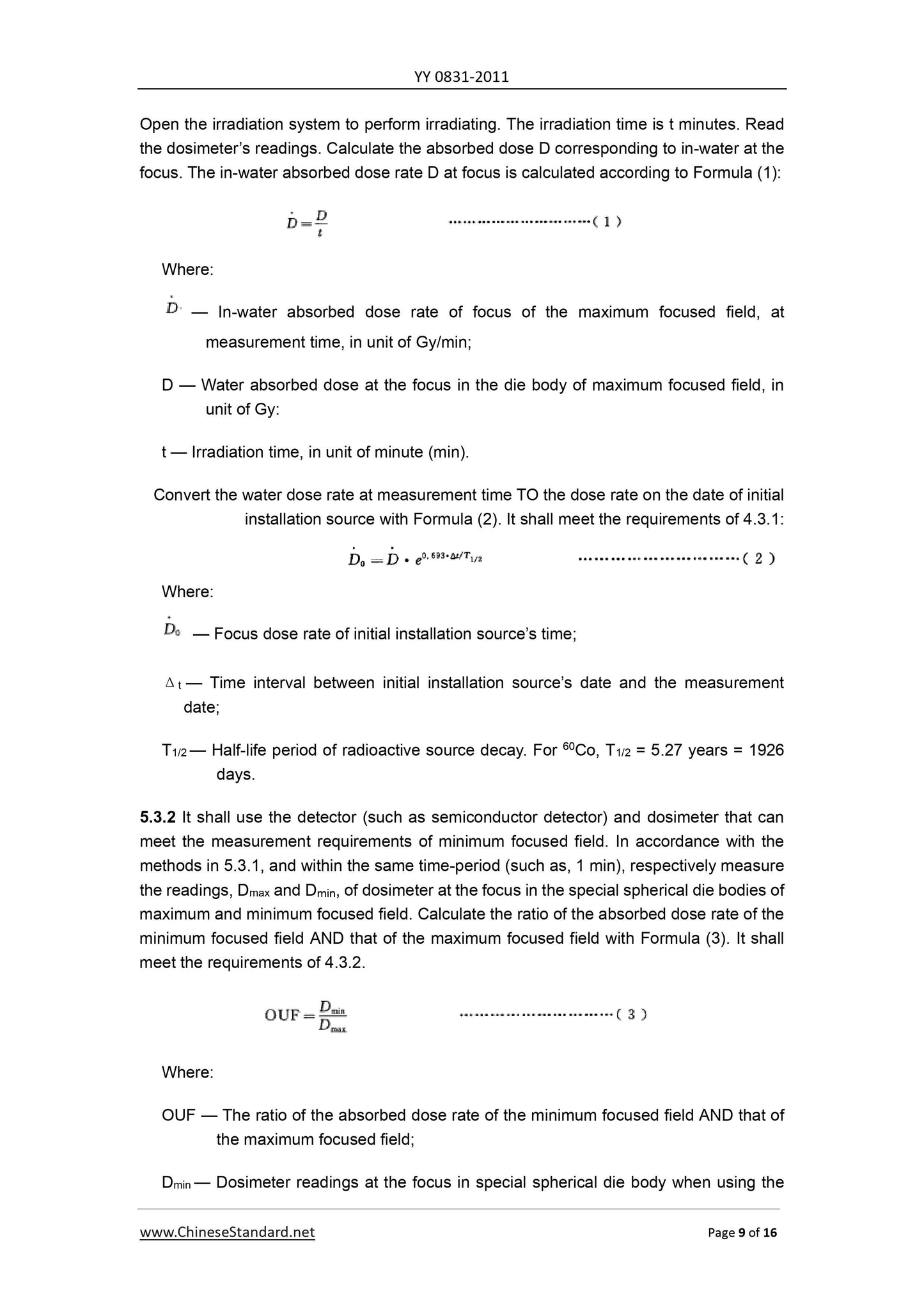
minimum focused field;
Dmax — Dosimeter readings at the focus in special spherical die body when using the
maximum focused field.
5.4 Focused field size
5.4.1 Grayscale-dose calibration
Radiate a series of films in the allowed test area of grayscale-dose response curve of the
film. Use a suitable mathematical model according to the radiating dose values and the
corresponding film’s grayscale value, Draw the grayscale-dose response curve for the
radiation films used.
5.4.2 Focused field film radiation
According to following steps to respectively measure the maximum deviations of the
focused field size and the nominal value.
a) Select a set of collimator for therapy. Measure at least on 2 focal planes;
b) Put the film of which the size fits the measurement of focused field onto the central
position of special die body. Let the film to be located on some focal plane;
c) Transfer the special die body, together with the therapeutic bed, into focus position.
Start to radiate. Radiation dose shall be within the appropriate response area of the
grayscale-dose response curve of the film used.
d) Repeat b). Let the film to be located on another focal plane. Repeat the radiation
operation of c);
e) Replace the collimator. Repeat tests b)-e). Complete the radiation of each group of
focused field;
f) Perform the therapy to the film.
5.4.3 Calculation of focused field size
According to following steps to calculate the focused field size.
a) Use a scanner to scan the film. The resolution shall not be less than 300 DPI.
Respectively scan the focused field films of 5.4.2;
b) Use the grayscale-dose response curve of 5.4.1 to convert the film grayscale TO
corresponding dose values. Draw a dose distribution curve on directions of 3 axes.
c) Separately find out the dose of each film at positioning reference point. Define the
dose value as the 100% dose value of the film;
planning software AND the ac...
Get QUOTATION in 1-minute: Click YY 0831.1-2011
Historical versions: YY 0831.1-2011
Preview True-PDF (Reload/Scroll if blank)
YY 0831.1-2011: Stereotactic radiotherapy system with gamma beam - Part 1: Multi-source stereotactic radiotherapy system with gamma beam for head lesion
YY 0831‐2011
YY
ICS 11.040.50
C 43
PHARMACEUTICAL INDUSTRY STANDARD
OF THE PEOPLE’S REPUBLIC OF CHINA
Stereotactic radiotherapy system with gamma
beam — Part 1. Multi-source stereotactic
radiotherapy system with gamma beam for head
lesion
ISSUED ON. DECEMBER 31, 2011
IMPLEMENTED ON. JUNE 1, 2013
Issued by. State Food and Drug Administration
Table of Contents
Foreword ... 3
1 Scope ... 4
2 Normative references ... 4
3 Terms and definitions ... 5
4 Requirements ... 5
5 Test methods ... 8
Annex A (Informative) Materials, structure and size of special spherical die
body and special focus measuring tools for test ... 15
Foreword
All technical contents of this Part are mandatory.
YY 0831 is consisted of 2 parts as follows.
— Part 1. Multi-source stereotactic radiotherapy system with gamma beam for head
lesion;
— Part 2. Multi-source stereotactic radiotherapy system with gamma beam for body
lesion.
This Part is part 1 of YY 0831.
This Part is drafted according to the rules specified in GB/T 1.1-2009.
Please note that some contents in this Document may involve patents. The issuing
authority of this Document does not undertake the responsibilities to identify these
patents.
The part was proposed by China Food and Drug Administration.
The part shall be under the jurisdiction of National Standardization Technical Committee of
Medical Appliances Radiotherapy and Technical Committee of Nuclear Medicine and
Radiation Dosimetry Equipment (SAC/TC10/SC3).
Drafting organization of this Part. Beijing Institute of Medical Device Testing, Shenzhen
Haibo Technology Co., Ltd., Medical Science and Technology Development (Shenzhen) Co.,
Ltd. (MASEP), Shenzhen Shengai Medical Technology Development Co., Ltd., and
Shenzhen Zunrui Technology Co., Ltd.
The main drafters of the part. Zhang Zhaoyuan, Zhang Xin, Song Lianyou, Chen Jing, Zhu
Weiqun, Xu Tao, Zheng Tie, and Liu Guangwu.
Stereotactic radiotherapy system with gamma beam —
Part 1. Multi-source stereotactic radiotherapy system
with gamma beam for head lesion
1 Scope
This Part of YY 0831 specifies the scope, terms, definitions, requirements, and test
methods of the multi-source stereotactic radiotherapy system with gamma beam for head
lesion.
This Part applies to the multi-source stereotactic radiotherapy system with gamma beam
for head lesion (hereinafter referred to as the system). The system simultaneously uses
multiple 60Co sealed radioactive sources (it may be dynamic or static) to conduct beaming
irradiation to the head lesion.
2 Normative references
The articles contained in the following documents have become part of this Document
when they are quoted herein. For the dated documents so quoted, all the modifications
(Including all corrections) or revisions made thereafter shall be applicable to this
Document.
GB 9706.1-2007 Medical electrical equipment - Part 1. General requirements for safety
GB 9706.15-2008 Medical electrical equipment - Part 1. General requirements for safety
- Collateral standard. Safety requirements for medical electrical systems
GB 9706.17-2009 Medical electrical equipment - Part 2. Particular requirements for the
safety of gamma beam therapy equipment
GB/T 17857-1999 Medical radiology – Terminology (Equipment for radiotherapy, nuclear
medicine and radiation dosimetry)
GB/T 18987-2003 Radiotherapy equipment - Coordinates, movements and scales
YY 0637-2012 Medical electrical equipment - Requirements for the safety of
radiotherapy therapeutic planning systems
c) For the focused field of which the nominal size is greater than 20 mm and less than
or equal to 30 mm, the dose gradient of each focused field shall not exceed 10 mm.
4.6 Deviation of positioning reference point
Deviation of the positioning reference point of the system shall meet the following
requirements.
a) For the minimum focused field, the deviation between the positioning reference point
and the focus shall be less than or equal to 0.5 mm;
b) The deviations of positioning reference point for each focused field shall be included
in accompanying documents.
4.7 Integrated error of dose calculation
For each focused field, the error between the absorbent dose value calculated by the
therapeutic planning software AND the actual measured value shall not exceed ±5%.
4.8 Position error of therapeutic plan reference point
Reconstruction position error of therapeutic planning software shall not exceed 1.5 mm.
4.9 Safety requirements
The system shall meet the requirements of GB 9706.1-2007, GB 9706.15-2008, GB
9706.17-2009 and YY 0637-2012.
5 Test methods
5.1 Coordinate system
Check the accompanying documents. It shall comply with the requirements of 4.1.
5.2 Accompanying documents
Check the accompanying documents. It shall comply with the requirements of 4.2.
5.3 Nominal absorbed dose rate of the focus
5.3.1 The ionization chamber and the dosimeter used for measurement shall meet the
requirements of the measurement conditions. Install the special spherical die body
(hereinafter referred to as "Special Die body", see Appendix A) of which the diameter is
160 mm on the positioning bracket. The die body’s center is located at the positioning
reference point. Insert it into ionization chamber, so as to make its effective measurement
point to coincide with the die body’s center. Transfer the special die body along with the
therapeutic bed into a predetermined irradiation position. Use the maximum focused field.
minimum focused field;
Dmax — Dosimeter readings at the focus in special spherical die body when using the
maximum focused field.
5.4 Focused field size
5.4.1 Grayscale-dose calibration
Radiate a series of films in the allowed test area of grayscale-dose response curve of the
film. Use a suitable mathematical model according to the radiating dose values and the
corresponding film’s grayscale value, Draw the grayscale-dose response curve for the
radiation films used.
5.4.2 Focused field film radiation
According to following steps to respectively measure the maximum deviations of the
focused field size and the nominal value.
a) Select a set of collimator for therapy. Measure at least on 2 focal planes;
b) Put the film of which the size fits the measurement of focused field onto the central
position of special die body. Let the film to be located on some focal plane;
c) Transfer the special die body, together with the therapeutic bed, into focus position.
Start to radiate. Radiation dose shall be within the appropriate response area of the
grayscale-dose response curve of the film used.
d) Repeat b). Let the film to be located on another focal plane. Repeat the radiation
operation of c);
e) Replace the collimator. Repeat tests b)-e). Complete the radiation of each group of
focused field;
f) Perform the therapy to the film.
5.4.3 Calculation of focused field size
According to following steps to calculate the focused field size.
a) Use a scanner to scan the film. The resolution shall not be less than 300 DPI.
Respectively scan the focused field films of 5.4.2;
b) Use the grayscale-dose response curve of 5.4.1 to convert the film grayscale TO
corresponding dose values. Draw a dose distribution curve on directions of 3 axes.
c) Separately find out the dose of each film at positioning reference point. Define the
dose value as the 100% dose value of the film;
planning software AND the ac...
Share