1
/
의
6
PayPal, credit cards. Download editable-PDF and invoice in 1 second!
YY/T 1465.3-2016 English PDF (YYT1465.3-2016)
YY/T 1465.3-2016 English PDF (YYT1465.3-2016)
정가
$150.00 USD
정가
할인가
$150.00 USD
단가
/
단위
배송료는 결제 시 계산됩니다.
픽업 사용 가능 여부를 로드할 수 없습니다.
Delivery: 3 seconds. Download true-PDF + Invoice.
Get QUOTATION in 1-minute: Click YY/T 1465.3-2016
Historical versions: YY/T 1465.3-2016
Preview True-PDF (Reload/Scroll if blank)
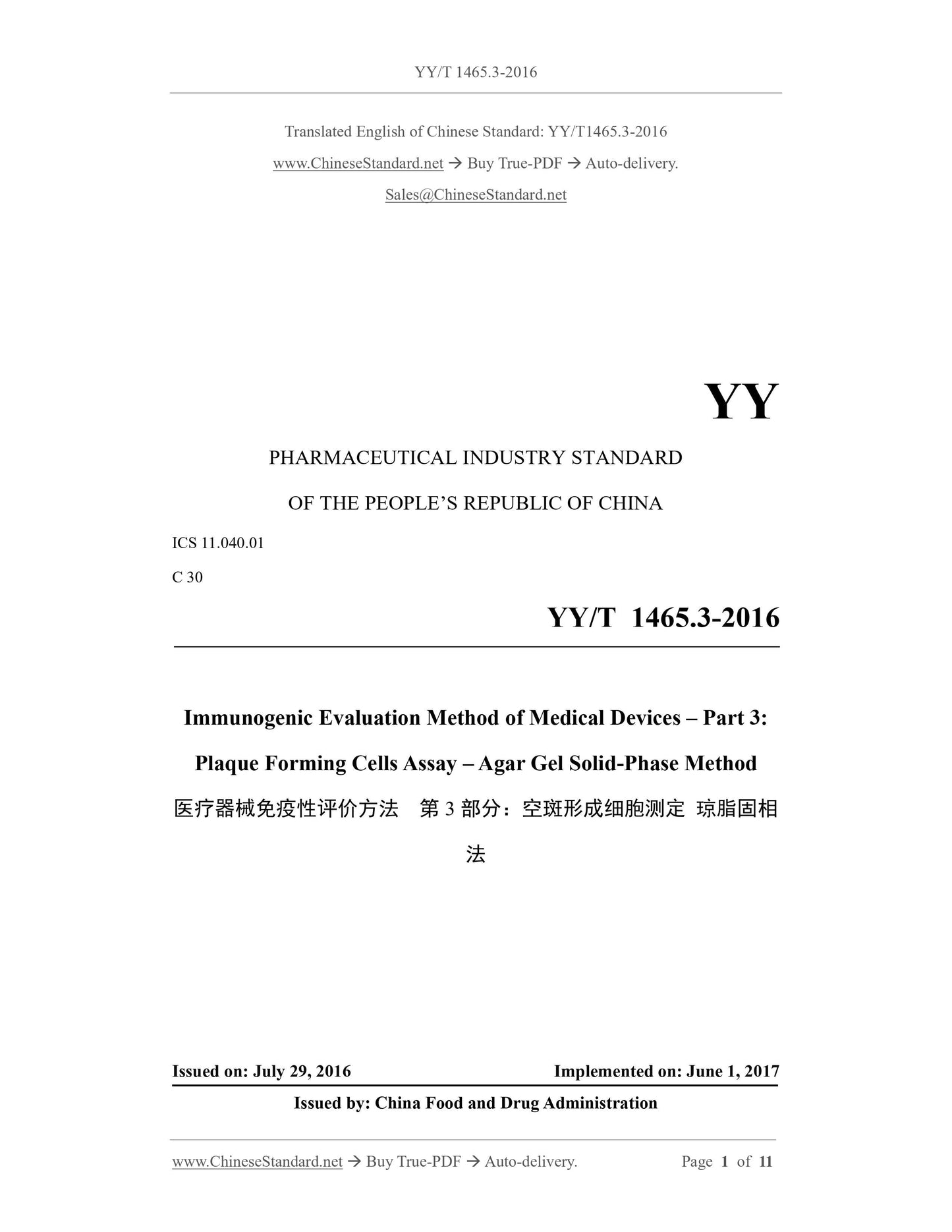
YY/T 1465.3-2016: Immunogenic evaluation method of medical devices--Part 3: Plaque forming cells assay--Agar gel solid-phase method
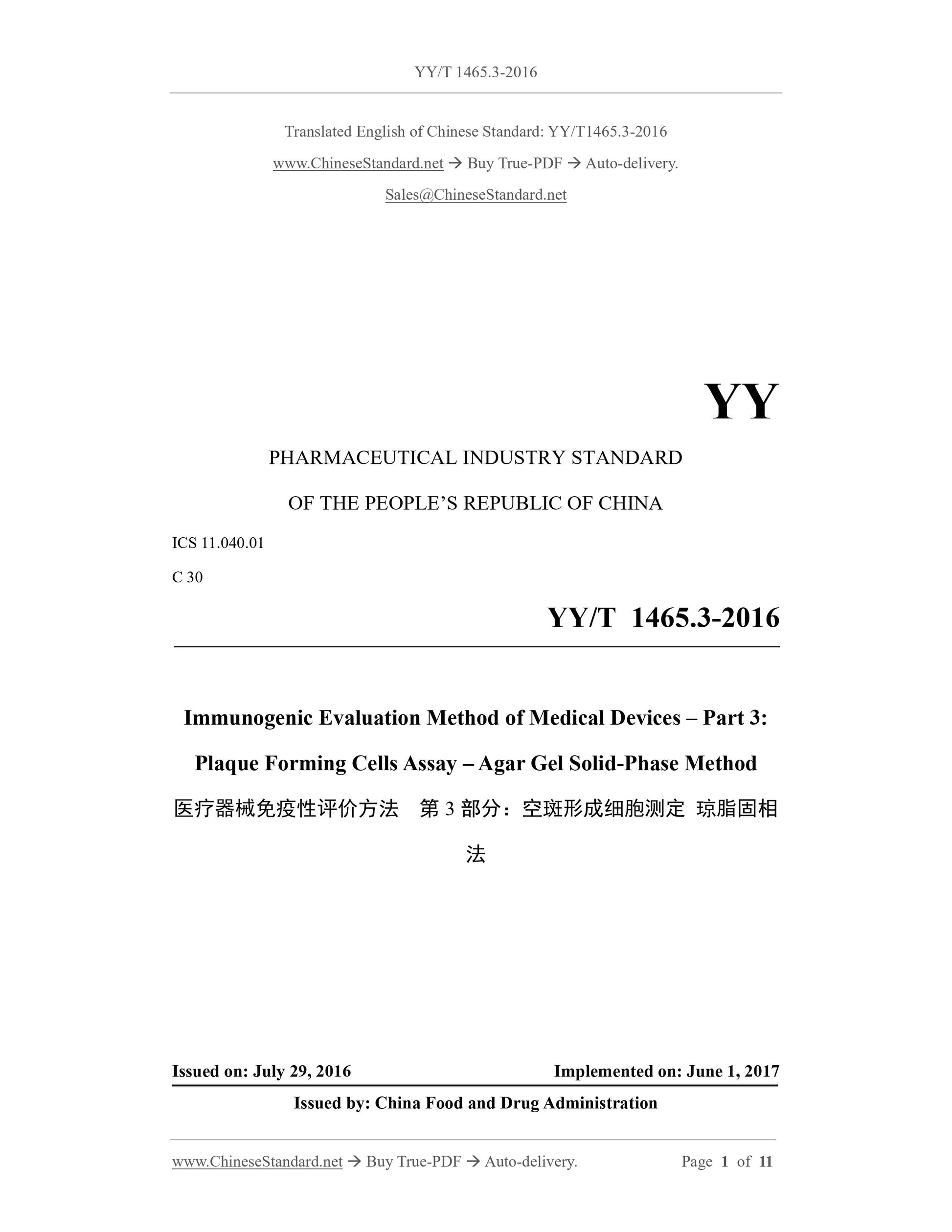
YY/T 1465.3-2016
YY
PHARMACEUTICAL INDUSTRY STANDARD
OF THE PEOPLE’S REPUBLIC OF CHINA
ICS 11.040.01
C 30
Immunogenic Evaluation Method of Medical Devices – Part 3:
Plaque Forming Cells Assay – Agar Gel Solid-Phase Method
ISSUED ON: JULY 29, 2016
IMPLEMENTED ON: JUNE 1, 2017
Issued by: China Food and Drug Administration
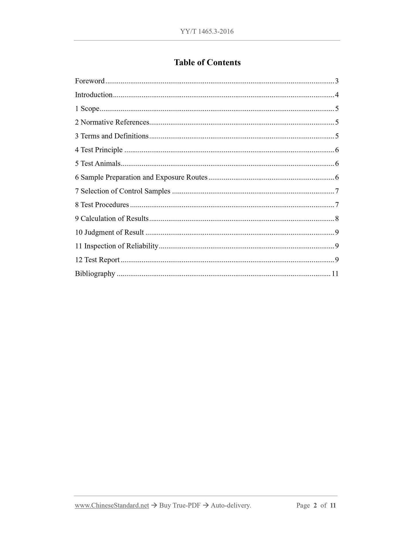
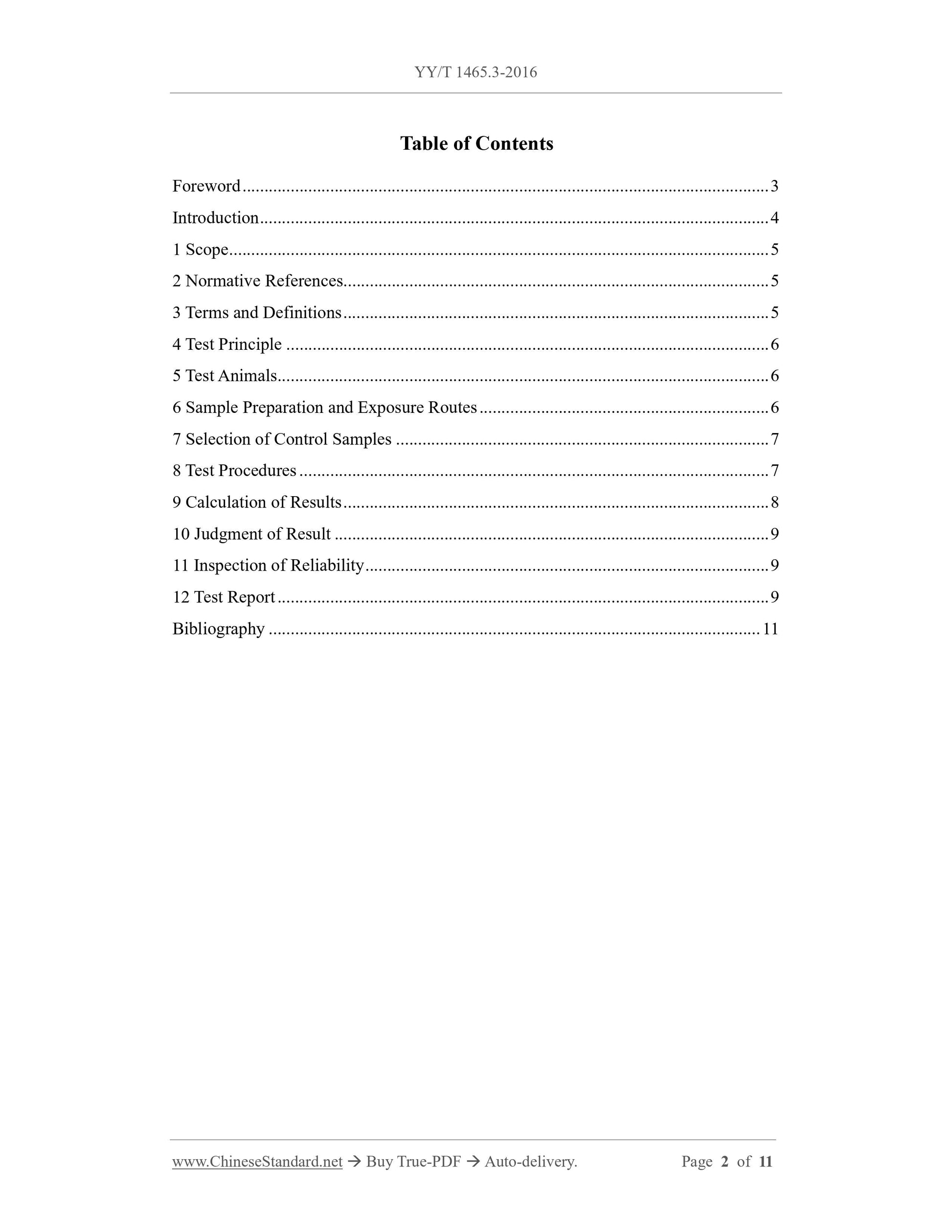
Table of Contents
Foreword ... 3
Introduction ... 4
1 Scope ... 5
2 Normative References ... 5
3 Terms and Definitions ... 5
4 Test Principle ... 6
5 Test Animals ... 6
6 Sample Preparation and Exposure Routes ... 6
7 Selection of Control Samples ... 7
8 Test Procedures ... 7
9 Calculation of Results ... 8
10 Judgment of Result ... 9
11 Inspection of Reliability ... 9
12 Test Report ... 9
Bibliography ... 11
Immunogenic Evaluation Method of Medical Devices – Part 3:
Plaque Forming Cells Assay – Agar Gel Solid-Phase Method
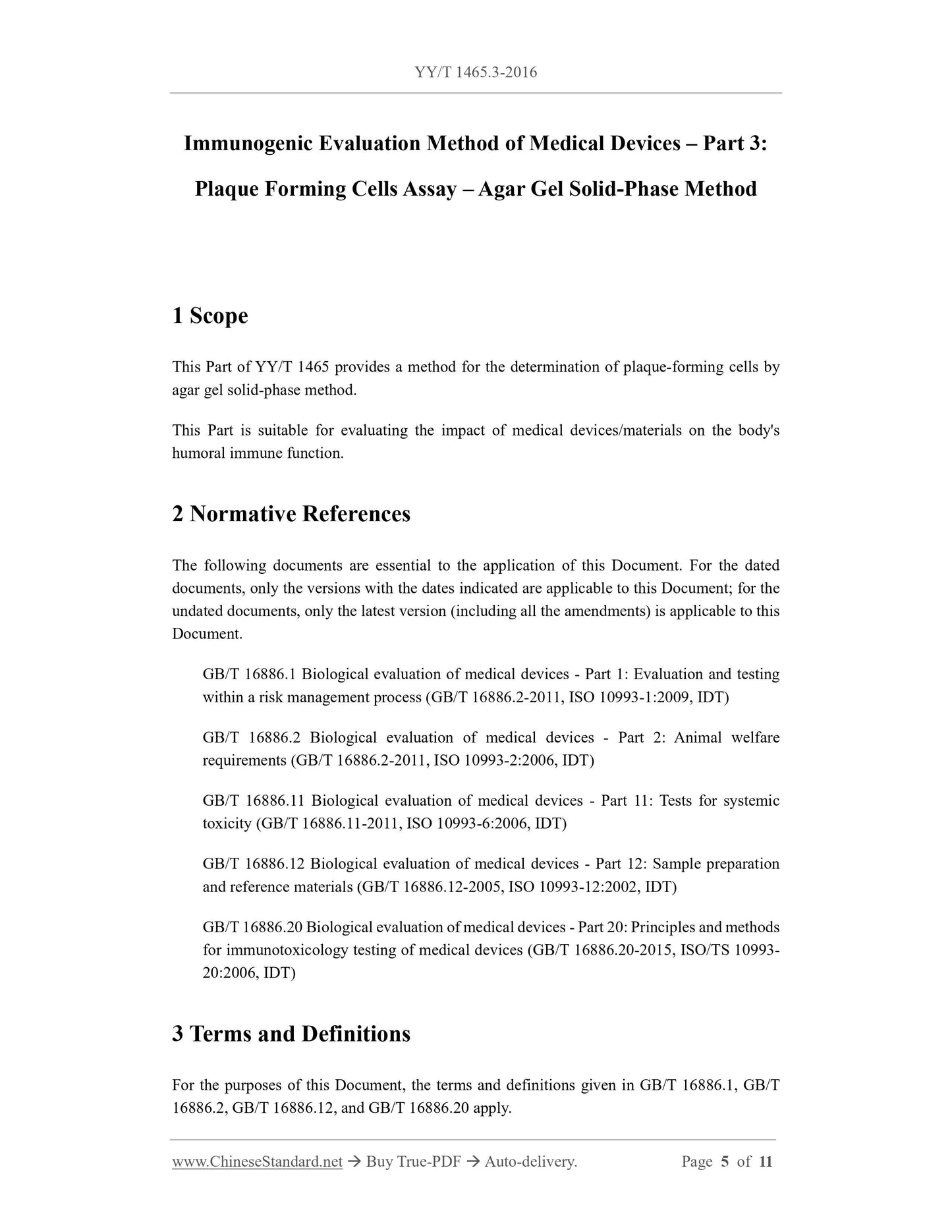
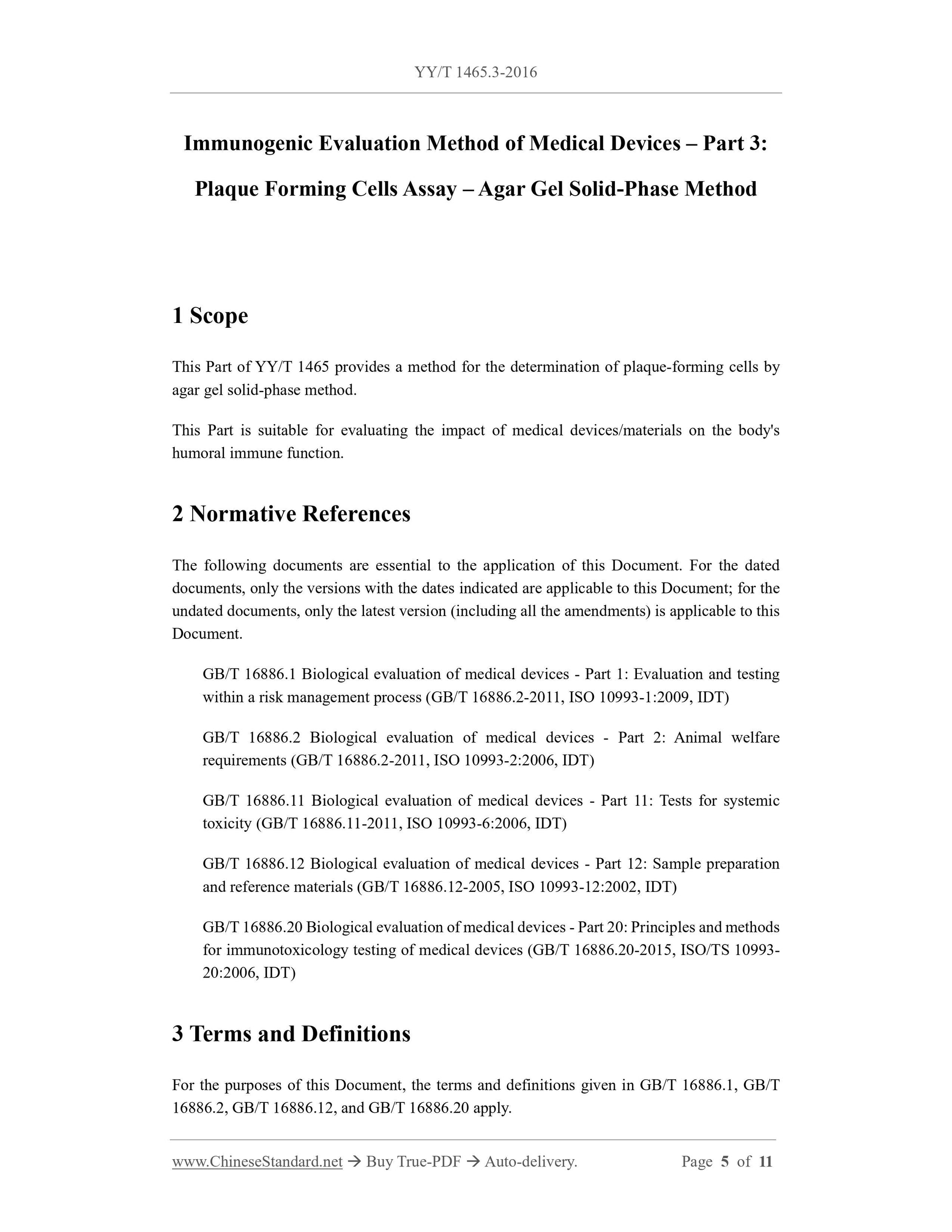
1 Scope
This Part of YY/T 1465 provides a method for the determination of plaque-forming cells by
agar gel solid-phase method.
This Part is suitable for evaluating the impact of medical devices/materials on the body's
humoral immune function.
2 Normative References
The following documents are essential to the application of this Document. For the dated
documents, only the versions with the dates indicated are applicable to this Document; for the
undated documents, only the latest version (including all the amendments) is applicable to this
Document.
GB/T 16886.1 Biological evaluation of medical devices - Part 1: Evaluation and testing
within a risk management process (GB/T 16886.2-2011, ISO 10993-1:2009, IDT)
GB/T 16886.2 Biological evaluation of medical devices - Part 2: Animal welfare
requirements (GB/T 16886.2-2011, ISO 10993-2:2006, IDT)
GB/T 16886.11 Biological evaluation of medical devices - Part 11: Tests for systemic
toxicity (GB/T 16886.11-2011, ISO 10993-6:2006, IDT)
GB/T 16886.12 Biological evaluation of medical devices - Part 12: Sample preparation
and reference materials (GB/T 16886.12-2005, ISO 10993-12:2002, IDT)
GB/T 16886.20 Biological evaluation of medical devices - Part 20: Principles and methods
for immunotoxicology testing of medical devices (GB/T 16886.20-2015, ISO/TS 10993-
20:2006, IDT)
3 Terms and Definitions
For the purposes of this Document, the terms and definitions given in GB/T 16886.1, GB/T
16886.2, GB/T 16886.12, and GB/T 16886.20 apply.
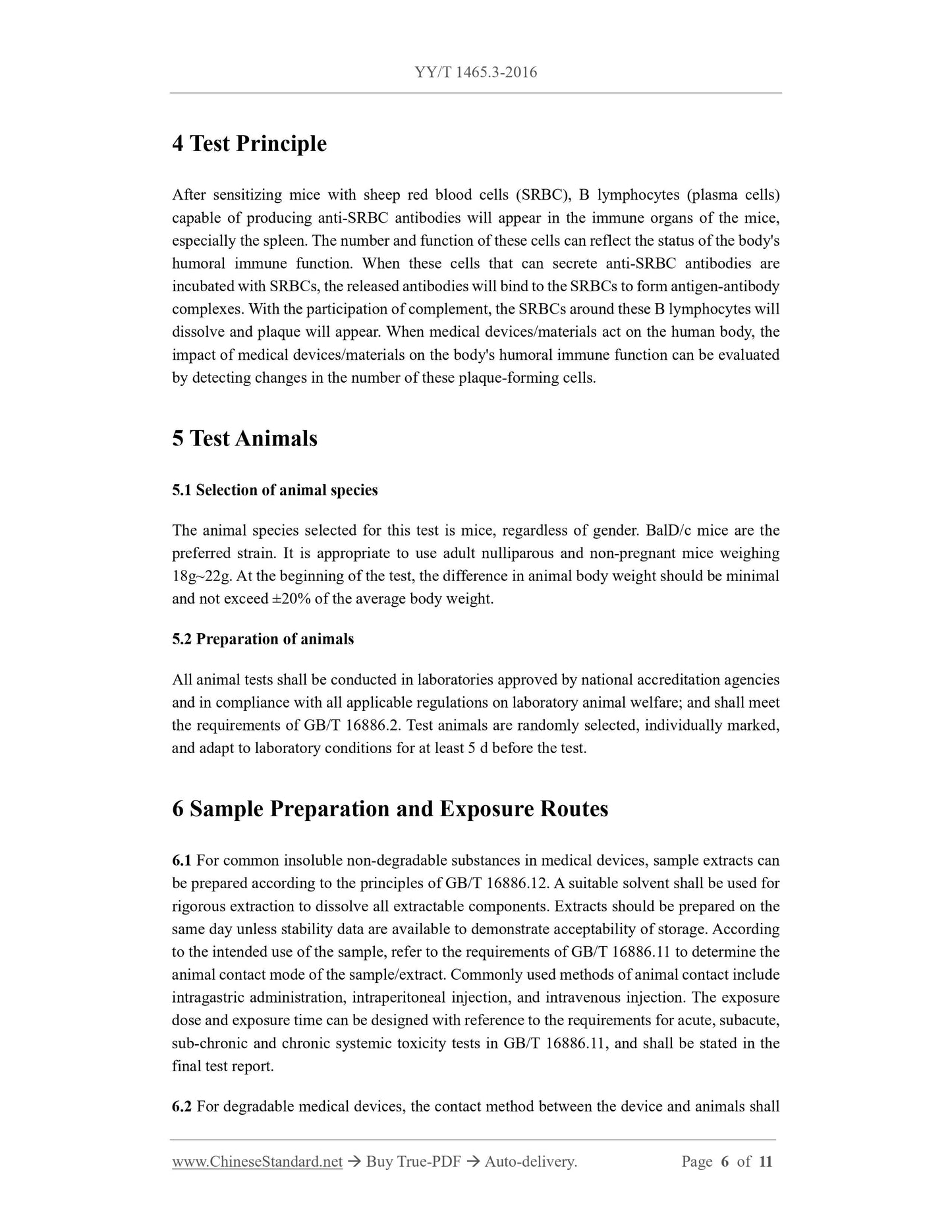
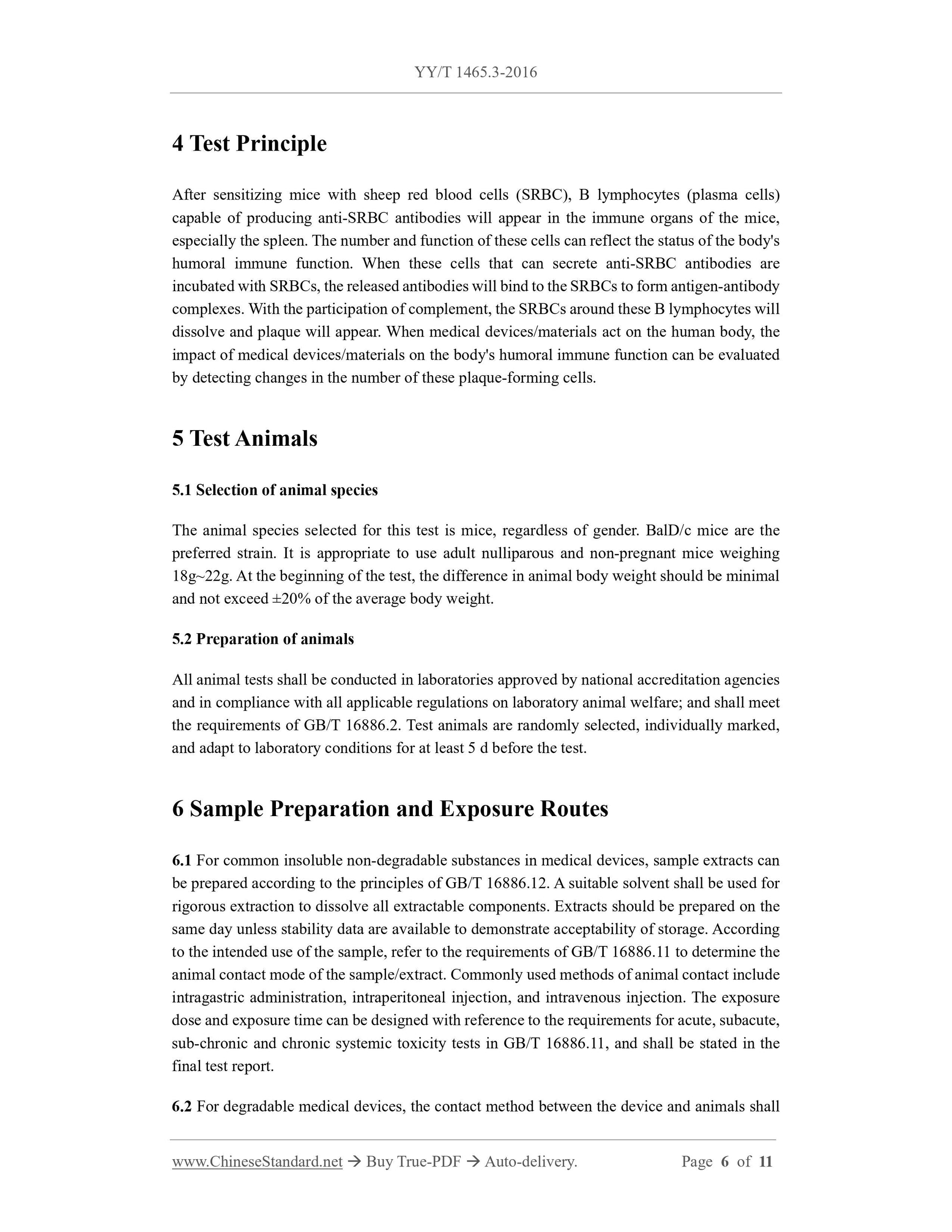
4 Test Principle
After sensitizing mice with sheep red blood cells (SRBC), B lymphocytes (plasma cells)
capable of producing anti-SRBC antibodies will appear in the immune organs of the mice,
especially the spleen. The number and function of these cells can reflect the status of the body's
humoral immune function. When these cells that can secrete anti-SRBC antibodies are
incubated with SRBCs, the released antibodies will bind to the SRBCs to form antigen-antibody
complexes. With the participation of complement, the SRBCs around these B lymphocytes will
dissolve and plaque will appear. When medical devices/materials act on the human body, the
impact of medical devices/materials on the body's humoral immune function can be evaluated
by detecting changes in the number of these plaque-forming cells.
5 Test Animals
5.1 Selection of animal species
The animal species selected for this test is mice, regardless of gender. BalD/c mice are the
preferred strain. It is appropriate to use adult nulliparous and non-pregnant mice weighing
18g~22g. At the beginning of the test, the difference in animal body weight should be minimal
and not exceed ±20% of the average body weight.
5.2 Preparation of animals
All animal tests shall be conducted in laboratories approved by national accreditation agencies
and in compliance with all applicable regulations on laboratory animal welfare; and shall meet
the requirements of GB/T 16886.2. Test animals are randomly selected, individually marked,
and adapt to laboratory conditions for at least 5 d before the test.
6 Sample Preparation and Exposure Routes
6.1 For common insoluble non-degradable substances in medical devices, sample extracts can
be prepared according to the principles of GB/T 16886.12. A suitable solvent shall be used for
rigorous extraction to dissolve all extractable components. Extracts should be prepared on the
same day unless stability data are available to demonstrate acceptability of storage. According
to the intended use of the sample, refer to the requirements of GB/T 16886.11 to determine the
animal contact mode of the sample/extract. Commonly used methods of animal contact include
intragastric administration, intraperitoneal injection, and intravenous injection. The exposure
dose and exposure time can be designed with reference to the requirements for acute, subacute,
sub-chronic and chronic systemic toxicity tests in GB/T 16886.11, and shall be stated in the
final test report.
6.2 For degradable medical devices, the contact method between the device and animals shall
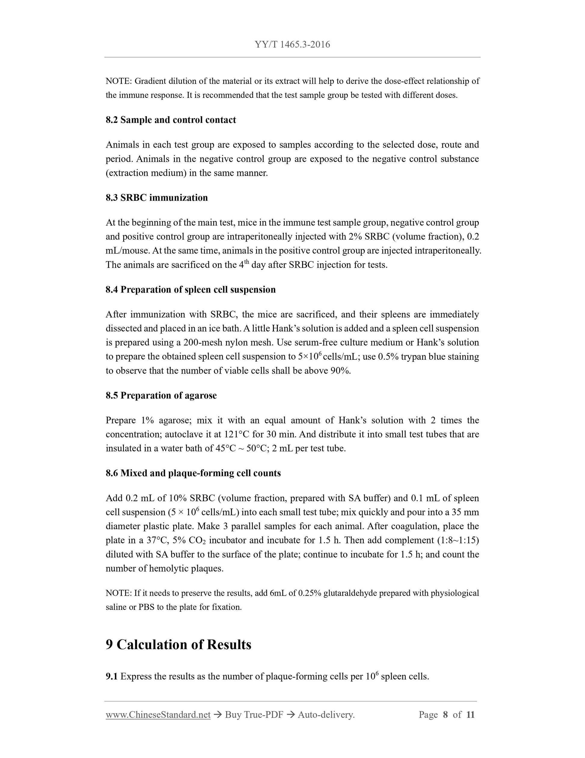
NOTE: Gradient dilution of the material or its extract will help to derive the dose-effect relationship of
the immune response. It is recommended that the test sample group be tested with different doses.
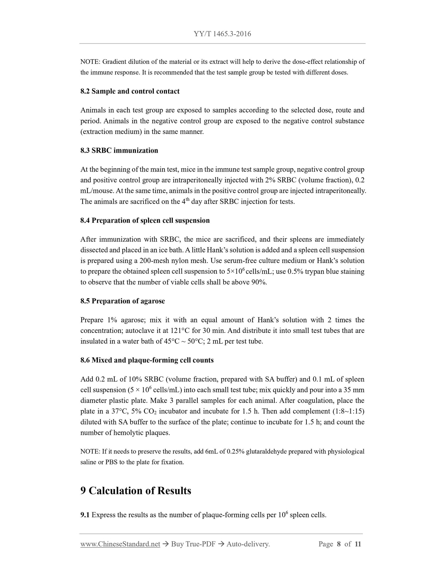
8.2 Sample and control contact
Animals in each test group are exposed to samples according to the selected dose, route and
period. Animals in the negative control group are exposed to the negative control substance
(extraction medium) in the same manner.
8.3 SRBC immunization
At the beginning of the main test, mice in the immune test sample group, negative control group
and positive control group are intraperitoneally injected with 2% SRBC (volume fraction), 0.2
mL/mouse. At the same time, animals in the positive control group are injected intraperitoneally.
The animals are sacrificed on the 4th day after SRBC injection for tests.
8.4 Preparation of spleen cell suspension
After immunization with SRBC, the mice are sacrificed, and their spleens are immediately
dissected and placed in an ice bath. A little Hank’s solution is added and a spleen cell suspension
is prepared using a 200-mesh nylon mesh. Use serum-free culture medium or Hank’s solution
to prepare the obtained spleen cell suspension to 5×10⁶ cells/mL; use 0.5% trypan blue staining
to observe that the number of viable cells shall be above 90%.
8.5 Preparation of agarose
Prepare 1% agarose; mix it with an equal amount of Hank’s solution with 2 times the
concentration; autoclave it at 121°C for 30 min. And distribute it into small test tubes that are
insulated in a water bath of 45°C ~ 50°C; 2 mL per test tube.
8.6 Mixed and plaque-forming cell counts
Add 0.2 mL of 10% SRBC (volume fraction, prepared with SA buffer) and 0.1 mL of spleen
cell suspension (5 × 10⁶ cells/mL) into each small test tube; mix quickly and pour into a 35 mm
diameter plastic plate. Make 3 parallel samples for each animal. After coagulation, place the
plate in a 37°C, 5% CO2 incubator and incubate for 1.5 h. Then add complement (1:8~1:15)
diluted with SA buffer to the surface of the plate; continue to incubate for 1.5 h; and count the
number of hemolytic plaques.
NOTE: If it needs to preserve the results, add 6mL of 0.25% glutaraldehyde prepared with physiological
saline or PBS to the plate for fixation.
9 Calculation of Results
9.1 Express the results as the number of plaque-forming cells per 106 spleen cells.
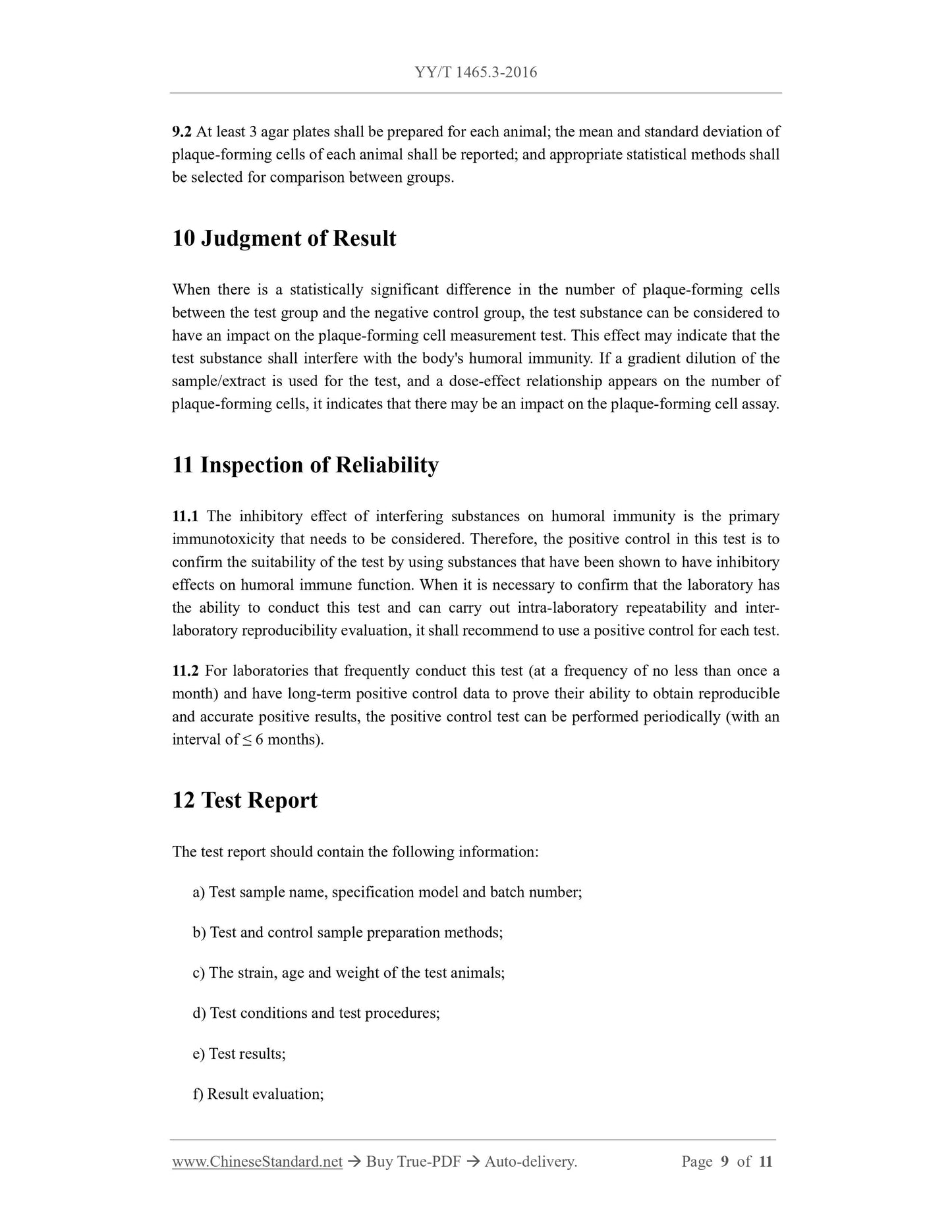
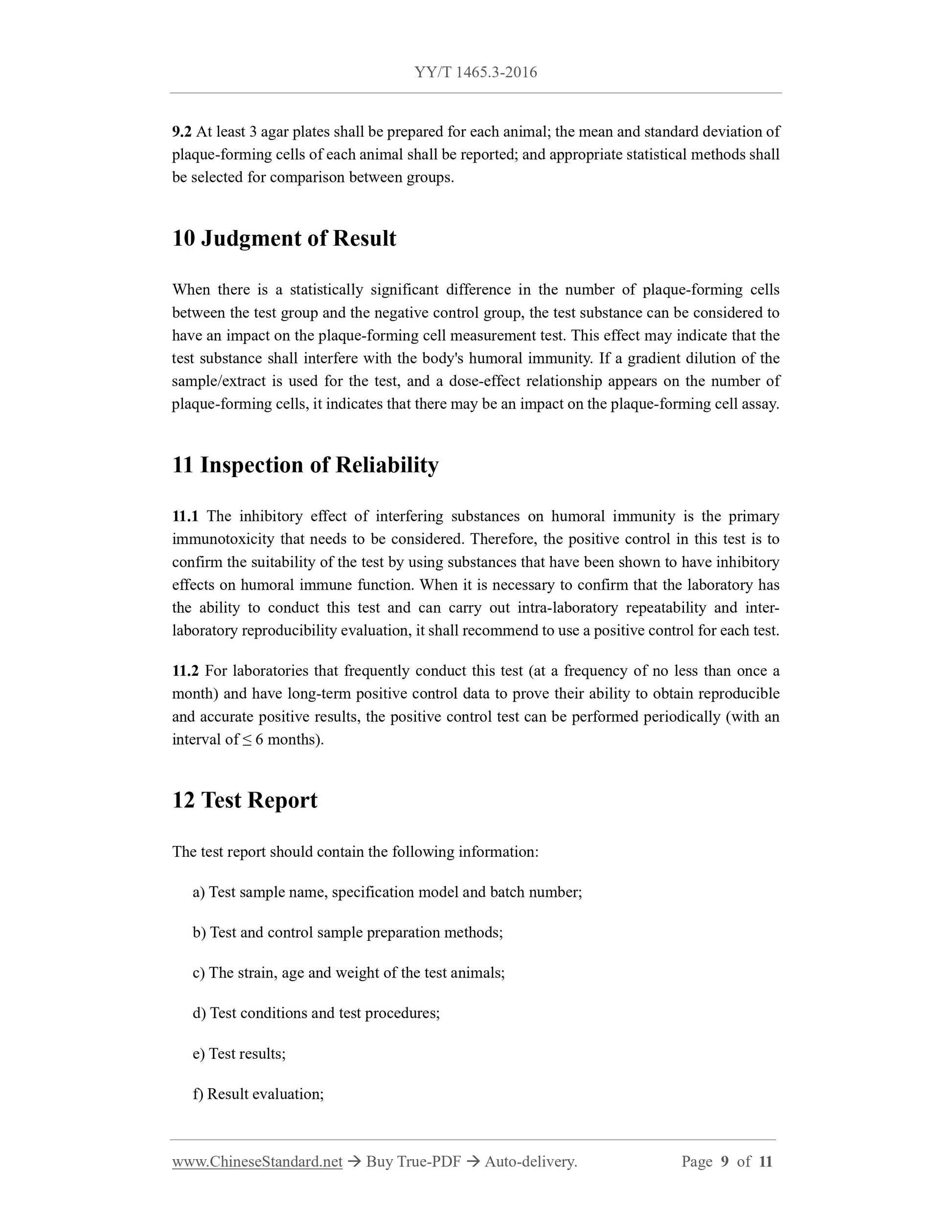
9.2 At least 3 agar plates shall be prepared for each animal; the mean and standard deviation of
plaque-forming cells of...
Share
