
1
/
의
9
PayPal, credit cards. Download editable-PDF and invoice in 1 second!
YY/T 1465.5-2016 English PDF (YYT1465.5-2016)
YY/T 1465.5-2016 English PDF (YYT1465.5-2016)
정가
$210.00 USD
정가
할인가
$210.00 USD
단가
/
단위
배송료는 결제 시 계산됩니다.
픽업 사용 가능 여부를 로드할 수 없습니다.
Delivery: 3 seconds. Download true-PDF + Invoice.
Get QUOTATION in 1-minute: Click YY/T 1465.5-2016
Historical versions: YY/T 1465.5-2016
Preview True-PDF (Reload/Scroll if blank)
YY/T 1465.5-2016: Immunogenic evaluation method of medical devices--Part 5: Determination of α-Gal antigen clearance in medical devices utilizing animal tissues and their derivatives with M86 antibody
YY/T 1465.5-2016
YY
PHARMACEUTICAL INDUSTRY STANDARD
OF THE PEOPLE’S REPUBLIC OF CHINA
ICS 11.120.20
C 30
Immunogenic evaluation method of medical devices - Part 5:
Determination of α-Gal antigen clearance in medical devices
utilizing animal tissues and their derivatives with M86
antibody
ISSUED ON: JULY 29, 2016
IMPLEMENTED ON: JUNE 01, 2017
Issued by: China Food and Drug Administration.
Table of Contents
Foreword ... 3
Introduction ... 4
1 General provisions ... 5
2 Normative references ... 5
3 Terms and definitions ... 6
4 Test principle ... 6
5 Reagents and instruments ... 6
6 Test steps ... 6
7 Data analysis ... 9
8 Test report ... 10
Appendix A (Informative) Example of determination of α-Gal antigen clearance rate in
enzymatically hydrolyzed pig skin products by immunohistochemistry ... 11
Appendix B (Informative) Example of sample processing ... 14
References ... 16
Immunogenic evaluation method of medical devices - Part 5:
Determination of α-Gal antigen clearance in medical devices
utilizing animal tissues and their derivatives with M86
antibody
1 General provisions
This Part of YY/T 1465 provides a method for determining the clearance rate of α-Gal
antigen in medical devices of animal origin, which is suitable for the evaluation of the
effectiveness of the α-Gal antigen clearance process.
The method in this Part is applicable to materials that can fully expose the α-Gal antigen
through grinding. If the α-Gal antigen cannot be fully exposed through grinding, other
confirmed suitable methods may be considered.
Note: Appendix A gives examples of immunohistochemistry evaluation methods.
2 Normative references
The following documents are essential to the application of this document. For the dated
documents, only the versions with the dates indicated are applicable to this document;
for the undated documents, only the latest version (including all the amendments) is
applicable to this standard.
GB/T 16886.2 Biological evaluation of medical devices - Part 2: Animal welfare
requirements (GB/T 16886.2-2011, ISO 10993-2:2006, IDT)
GB/T 16886.20 Biological evaluation of medical devices - Part 20: Principles and
methods for immunotoxicology testing of medical devices (GB/T 16886.20-2015,
ISO/TS 10993-20:2006, IDT)
YY/T 0771.1 Medical devices utilizing animal tissues and their derivatives - Part 1:
Application of risk management (YY/T 0771.1-2009, ISO 22442-1:2007, IDT)
Chinese Pharmacopoeia 2010 Edition
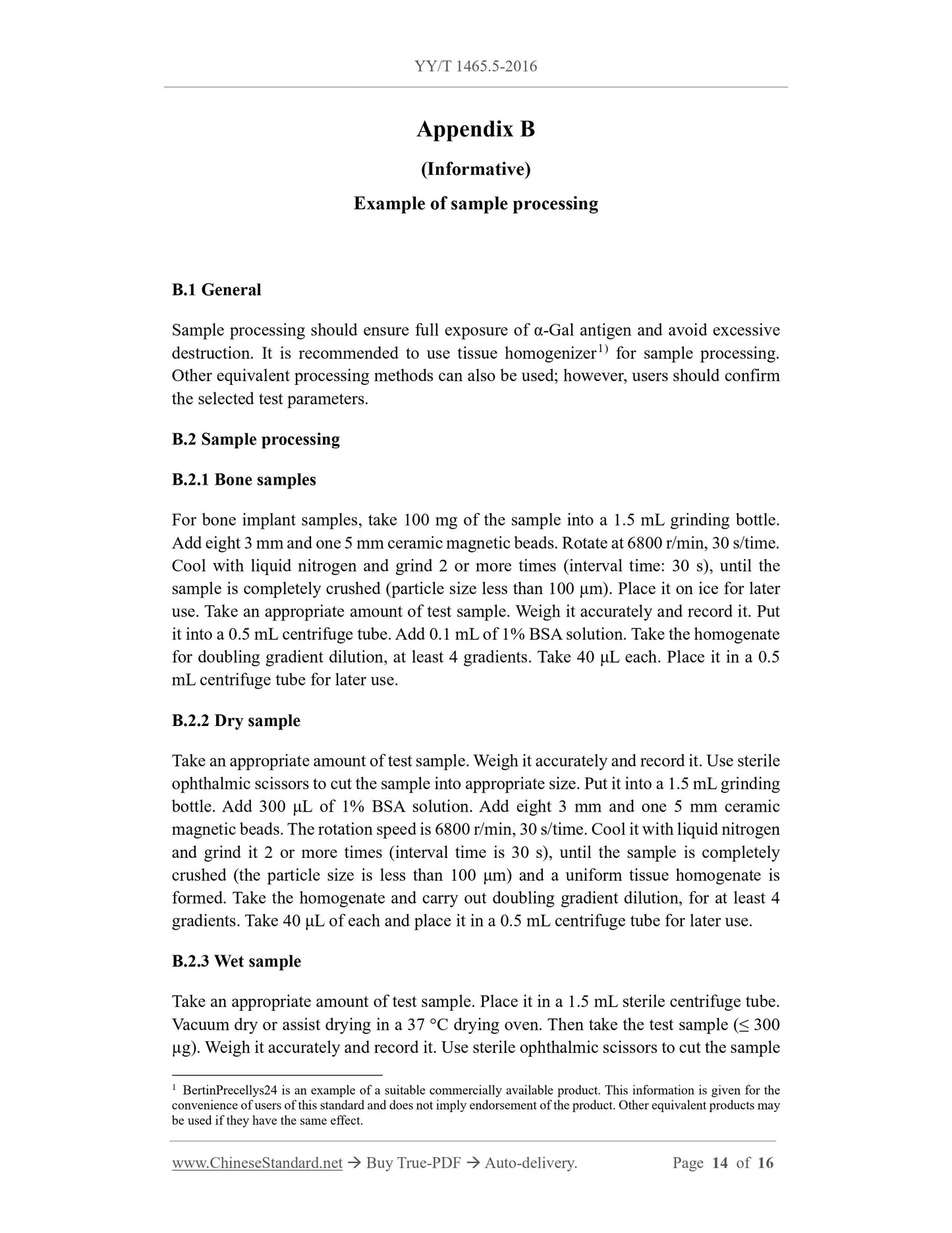
avoiding excessive destruction. The recommended particle size distribution range of
the processed sample is (0 μm ~ 90 μm). Examples of different types of sample
processing are given in Appendix B.
Note 1: Animal-derived biological materials are mainly composed of structural proteins, such
as collagen fibers. Experimenters should use appropriate sample preparation methods, to ensure
that the α-Gal antigen in the test sample is fully exposed.
Note 2: Three parallel samples are prepared for each sample; the average value is taken as the
final measurement result.
Note 3: According to the request of the client, 3 samples of different batch numbers or the same
batch number can be taken, to evaluate the process stability of different batches or the same
batch.
6.2 Preparation of test system control materials
6.2.1 General
Considering that the methods in this standard are affected by many factors, appropriate
control substances should be used, to verify the rationality of the test system before
testing. It is recommended to use mouse myeloma (SP2/0) cells or rabbit red blood cells
(RRBC). If other reference substances are selected, their effectiveness should be
confirmed.
6.2.2 SP2/0 cells
SP2/0 (ATCC CRL-1581TM) is obtained from the fusion of spleen B cells of BALB/c
mice immunized with sheep red blood cells and mouse myeloma cells. It does not
secrete immunoglobulins. Use RPMI1640 complete medium to culture, until the growth
state is good; adjust the cell concentration to 1 × 10⁷ cells/mL for later use.
6.2.3 RRBC
6.2.3.1 It is recommended to use untested healthy New Zealand rabbits, SPF grade. If
other strains of rabbits are selected, their suitability should be explained. Animals
should be kept for at least 5 days before blood collection, to adapt to the laboratory
environment. All animal testing should be conducted in laboratories approved by
national accreditation agencies and in compliance with all applicable regulations on
laboratory animal welfare, meanwhile it should also comply with the requirements of
GB/T 16886.2.
6.2.3.2 Take 20 mL of fresh rabbit whole blood. Add it to an Erlenmeyer flask
containing glass beads. Shake for 10 minutes, to remove fibrin. Add about 10 times the
amount of 0.9% sodium chloride solution. Shake well. Centrifuge at 2000 r/min for 5
minutes. Remove the supernatant. Wash the precipitated red blood cells 3 times, using
0.9% sodium chloride solution, according to the above method, until the supernatant is
no longer red. The obtained red blood cells are mixed with 0.9% sodium chloride
solution to 1 × 10⁷ cells/mL. Store it at 4 °C for later use.
6.2.4 Preparation steps
Resuspend 1 × 10⁶ SP2/0 cells or RRBC in 40 μL of 1% BSA. Mix well. Dilute 20 μL
of cells to create a total of 4 concentration gradients (including a blank group without
cells). Add 1% BSA solution to the cell stock solution and diluent, respectively, 80
μL/tube. Then the volume ratio of the highest concentration group containing the cell
stock solution is 20%. The volume ratio of the remaining groups decreases in a gradient
with the doubling dilution. Three replicate holes for each concentration gradient are
available for use.
6.3 Test grouping
Test grouping includes:
a) Material group without α-Gal antigen removal process;
b) Material group after α-Gal antigen removal process;
c) Negative control group, materials that do not contain α-Gal antigen;
d) Test solvent control group.
6.4 Determination of optimal M86 antibody dilution
6.4.1 General
There may be certain differences in the potency of different batches of M86 antibodies;
the optimal dilution of each batch of M86 antibodies should be determined. Divide the
commercially available M86 into appropriate volumes. Store it for later use.
6.4.2 Preparation of α-Gal antigen standard
Take α-Gal-BSA. Dissolve it in the coating solution at pH 9.6. Add it to the microplate,
100 μL/hole. The recommended final concentration is 1 μg/mL ~ 10 μg/mL. Coat
overnight at 4 °C. Use 1% BSA sealed for later use.
6.4.3 Optimal dilution of M86 antibody
Add 100 μL of diluted M86 antibody solution to each microplate, for a total of 8
dilutions. The recommended initial dilution is 1:25. The dilution is between 1:25 and
1:3200. Mix well. Incubate with shaking at room temperature for 1.5 hours. Use
washing solution to wash the plate three times. Add HRP-labeled anti-mouse secondary
antibody. Incubate with shaking at 37 °C for 1 hour. Then use washing solution to wash
the plate three times. After color development, use a microplate reader to read the
absorbance value. Draw a reaction curve between the absorbance value and the
corresponding M86 dilution. Take twice the concentration value of the dilution, that
just enters the plateau area of the reaction curve, as the optimal dilution. Dilute the
stored M86 antibody before each use.
6.5 M86 antibody co-incubation
Add 160 μL of the M86 antibody solution, at the dilut...
Get QUOTATION in 1-minute: Click YY/T 1465.5-2016
Historical versions: YY/T 1465.5-2016
Preview True-PDF (Reload/Scroll if blank)
YY/T 1465.5-2016: Immunogenic evaluation method of medical devices--Part 5: Determination of α-Gal antigen clearance in medical devices utilizing animal tissues and their derivatives with M86 antibody
YY/T 1465.5-2016
YY
PHARMACEUTICAL INDUSTRY STANDARD
OF THE PEOPLE’S REPUBLIC OF CHINA
ICS 11.120.20
C 30
Immunogenic evaluation method of medical devices - Part 5:
Determination of α-Gal antigen clearance in medical devices
utilizing animal tissues and their derivatives with M86
antibody
ISSUED ON: JULY 29, 2016
IMPLEMENTED ON: JUNE 01, 2017
Issued by: China Food and Drug Administration.
Table of Contents
Foreword ... 3
Introduction ... 4
1 General provisions ... 5
2 Normative references ... 5
3 Terms and definitions ... 6
4 Test principle ... 6
5 Reagents and instruments ... 6
6 Test steps ... 6
7 Data analysis ... 9
8 Test report ... 10
Appendix A (Informative) Example of determination of α-Gal antigen clearance rate in
enzymatically hydrolyzed pig skin products by immunohistochemistry ... 11
Appendix B (Informative) Example of sample processing ... 14
References ... 16
Immunogenic evaluation method of medical devices - Part 5:
Determination of α-Gal antigen clearance in medical devices
utilizing animal tissues and their derivatives with M86
antibody
1 General provisions
This Part of YY/T 1465 provides a method for determining the clearance rate of α-Gal
antigen in medical devices of animal origin, which is suitable for the evaluation of the
effectiveness of the α-Gal antigen clearance process.
The method in this Part is applicable to materials that can fully expose the α-Gal antigen
through grinding. If the α-Gal antigen cannot be fully exposed through grinding, other
confirmed suitable methods may be considered.
Note: Appendix A gives examples of immunohistochemistry evaluation methods.
2 Normative references
The following documents are essential to the application of this document. For the dated
documents, only the versions with the dates indicated are applicable to this document;
for the undated documents, only the latest version (including all the amendments) is
applicable to this standard.
GB/T 16886.2 Biological evaluation of medical devices - Part 2: Animal welfare
requirements (GB/T 16886.2-2011, ISO 10993-2:2006, IDT)
GB/T 16886.20 Biological evaluation of medical devices - Part 20: Principles and
methods for immunotoxicology testing of medical devices (GB/T 16886.20-2015,
ISO/TS 10993-20:2006, IDT)
YY/T 0771.1 Medical devices utilizing animal tissues and their derivatives - Part 1:
Application of risk management (YY/T 0771.1-2009, ISO 22442-1:2007, IDT)
Chinese Pharmacopoeia 2010 Edition
avoiding excessive destruction. The recommended particle size distribution range of
the processed sample is (0 μm ~ 90 μm). Examples of different types of sample
processing are given in Appendix B.
Note 1: Animal-derived biological materials are mainly composed of structural proteins, such
as collagen fibers. Experimenters should use appropriate sample preparation methods, to ensure
that the α-Gal antigen in the test sample is fully exposed.
Note 2: Three parallel samples are prepared for each sample; the average value is taken as the
final measurement result.
Note 3: According to the request of the client, 3 samples of different batch numbers or the same
batch number can be taken, to evaluate the process stability of different batches or the same
batch.
6.2 Preparation of test system control materials
6.2.1 General
Considering that the methods in this standard are affected by many factors, appropriate
control substances should be used, to verify the rationality of the test system before
testing. It is recommended to use mouse myeloma (SP2/0) cells or rabbit red blood cells
(RRBC). If other reference substances are selected, their effectiveness should be
confirmed.
6.2.2 SP2/0 cells
SP2/0 (ATCC CRL-1581TM) is obtained from the fusion of spleen B cells of BALB/c
mice immunized with sheep red blood cells and mouse myeloma cells. It does not
secrete immunoglobulins. Use RPMI1640 complete medium to culture, until the growth
state is good; adjust the cell concentration to 1 × 10⁷ cells/mL for later use.
6.2.3 RRBC
6.2.3.1 It is recommended to use untested healthy New Zealand rabbits, SPF grade. If
other strains of rabbits are selected, their suitability should be explained. Animals
should be kept for at least 5 days before blood collection, to adapt to the laboratory
environment. All animal testing should be conducted in laboratories approved by
national accreditation agencies and in compliance with all applicable regulations on
laboratory animal welfare, meanwhile it should also comply with the requirements of
GB/T 16886.2.
6.2.3.2 Take 20 mL of fresh rabbit whole blood. Add it to an Erlenmeyer flask
containing glass beads. Shake for 10 minutes, to remove fibrin. Add about 10 times the
amount of 0.9% sodium chloride solution. Shake well. Centrifuge at 2000 r/min for 5
minutes. Remove the supernatant. Wash the precipitated red blood cells 3 times, using
0.9% sodium chloride solution, according to the above method, until the supernatant is
no longer red. The obtained red blood cells are mixed with 0.9% sodium chloride
solution to 1 × 10⁷ cells/mL. Store it at 4 °C for later use.
6.2.4 Preparation steps
Resuspend 1 × 10⁶ SP2/0 cells or RRBC in 40 μL of 1% BSA. Mix well. Dilute 20 μL
of cells to create a total of 4 concentration gradients (including a blank group without
cells). Add 1% BSA solution to the cell stock solution and diluent, respectively, 80
μL/tube. Then the volume ratio of the highest concentration group containing the cell
stock solution is 20%. The volume ratio of the remaining groups decreases in a gradient
with the doubling dilution. Three replicate holes for each concentration gradient are
available for use.
6.3 Test grouping
Test grouping includes:
a) Material group without α-Gal antigen removal process;
b) Material group after α-Gal antigen removal process;
c) Negative control group, materials that do not contain α-Gal antigen;
d) Test solvent control group.
6.4 Determination of optimal M86 antibody dilution
6.4.1 General
There may be certain differences in the potency of different batches of M86 antibodies;
the optimal dilution of each batch of M86 antibodies should be determined. Divide the
commercially available M86 into appropriate volumes. Store it for later use.
6.4.2 Preparation of α-Gal antigen standard
Take α-Gal-BSA. Dissolve it in the coating solution at pH 9.6. Add it to the microplate,
100 μL/hole. The recommended final concentration is 1 μg/mL ~ 10 μg/mL. Coat
overnight at 4 °C. Use 1% BSA sealed for later use.
6.4.3 Optimal dilution of M86 antibody
Add 100 μL of diluted M86 antibody solution to each microplate, for a total of 8
dilutions. The recommended initial dilution is 1:25. The dilution is between 1:25 and
1:3200. Mix well. Incubate with shaking at room temperature for 1.5 hours. Use
washing solution to wash the plate three times. Add HRP-labeled anti-mouse secondary
antibody. Incubate with shaking at 37 °C for 1 hour. Then use washing solution to wash
the plate three times. After color development, use a microplate reader to read the
absorbance value. Draw a reaction curve between the absorbance value and the
corresponding M86 dilution. Take twice the concentration value of the dilution, that
just enters the plateau area of the reaction curve, as the optimal dilution. Dilute the
stored M86 antibody before each use.
6.5 M86 antibody co-incubation
Add 160 μL of the M86 antibody solution, at the dilut...
Share