


1
/
of
12
www.ChineseStandard.us -- Field Test Asia Pte. Ltd.
GB/T 13091-2018 English PDF (GB/T13091-2018)
GB/T 13091-2018 English PDF (GB/T13091-2018)
Regular price
$255.00
Regular price
Sale price
$255.00
Unit price
/
per
Shipping calculated at checkout.
Couldn't load pickup availability
GB/T 13091-2018: Determination of Salmonella in feeds
Delivery: 9 seconds. Download (& Email) true-PDF + Invoice.
Get Quotation: Click GB/T 13091-2018 (Self-service in 1-minute)
Historical versions (Master-website): GB/T 13091-2018
Preview True-PDF (Reload/Scroll-down if blank)
GB/T 13091-2018
GB
NATIONAL STANDARD OF THE
PEOPLE’S REPUBLIC OF CHINA
ICS 65.120
B 46
Replacing GB/T 13091-2002
Determination of Salmonella in feeds
ISSUED ON: SEPTEMBER 17, 2018
IMPLEMENTED ON: APRIL 01, 2019
Issued by: State Administration for Market Regulation;
Standardization Administration of PRC.
Table of Contents
Foreword ... 3
1 Scope ... 4
2 Normative references ... 4
3 Medium or material... 4
4 Instruments and equipment ... 5
5 Samples ... 5
6 Test method (see Appendix A for the test procedure diagram) ... 6
7 Expression of results ... 10
Appendix A (Normative) Test procedure diagram ... 11
Appendix B (Normative) Preparation and test methods of medium and reagents ... 12
Determination of Salmonella in feeds
1 Scope
This standard specifies the inspection methods for Salmonella in feed.
This standard applies to the inspection of Salmonella in feed and feed additives.
2 Normative references
The following documents are essential to the application of this document. For the dated
documents, only the versions with the dates indicated are applicable to this document;
for the undated documents, only the latest version (including all the amendments) is
applicable to this standard.
GB/T 6682 Water for analytical laboratory use - Specification and test methods
3 Medium or material
Unless otherwise stated, only reagents confirmed to be of analytical grade are used in
the analysis.
3.1 Water: It shall meet the requirements of grade-3 water in GB/T 6682.
3.2 Buffered peptone water (BPW): See B.1 in Appendix B.
3.3 Magnesium chloride malachite green (RV) enrichment solution: See B.2 in
Appendix B.
3.4 Cystine selenite (SC) enrichment solution: See B.3 in Appendix B.
3.5 Bismuth sulfite (BS) agar: See B.4 in Appendix B.
3.6 Deoxycholate-Hydrogen-Sulfide-Lactose (DHL) agar: See B.5 in Appendix B.
3.7 Salmonella chromogenic medium.
3.8 Nutrient agar (NA): See B.6 in Appendix B.
3.9 Trisaccharide iron agar (TSI): See B.7 in Appendix B.
3.10 Peptone water and indigo-based reagents: See B.8 in Appendix B.
3.11 Urea agar (pH7.2): See B.9 in Appendix B.
3.12 Potassium cyanide (KCN) medium: See B.10 in Appendix B.
3.13 Lysine decarboxylase test medium: See B.11 in Appendix B.
3.14 Sugar fermentation tube: See B.12 in Appendix B.
3.15 Ortho-nitrophenol β-D-galactopyranoside (ONPG) medium: See B.13 in
Appendix B.
3.16 Semi-solid agar: See B.14 in Appendix B.
3.17 Sodium malonate medium: See B.15 in Appendix B.
3.18 Salmonella factor O and H polyvalent diagnostic serum, Vi factor diagnostic serum.
4 Instruments and equipment
4.1 Refrigerator: 2 °C ~ 5 °C.
4.2 Constant temperature incubator: 36 °C ± 1 °C, 42 °C ± 1 °C.
4.3 Homogenizer.
4.4 Oscillators.
4.5 Electronic balance: Sensitivity 0.1 g.
4.6 Sterile conical flasks: Capacity 500 mL, 250 mL, or sterile homogeneous bag.
4.7 Sterile pipettes: 1 mL (with 0.01 mL scale), 10 mL (with 0.1 mL scale), or
micropipette and tip.
4.8 Sterile petri dishes: Diameter 60 mm, 90 mm.
4.9 Sterile test tubes: 3 mm × 50 mm, 10 mm × 75 mm.
4.10 pH meter or pH colorimetric tube or precision pH test paper.
4.11 Automatic microbial biochemical identification system.
5 Samples
5.1 Sampling principles
The collection of samples shall follow the principles of randomness and
representativeness. The sampling process shall follow aseptic procedures, to prevent all
possible external contamination.
5.2 Sampling method
5.2.1 Samples shall be collected from the same batch of products. The sampling volume
of each sample shall meet the requirements for microbiological index inspection, which
is generally not less than 500 g (mL).
5.2.2 For solid products not larger than 500 g or liquid products not larger than 500 mL
in independent packaging, take the complete package.
5.2.3 For individually packaged liquid products larger than 500 mL, it shall be shaken
or stirred with a sterile stick before sampling, to make them uniform, then collect an
appropriate amount of samples and put them into a sterile sampling container, as a
sample.
5.2.4 For solid products with individual packages greater than 500 g, use a sterile
sampler to take appropriate samples from different parts of the same package; put them
into the same sterile sampling container, as one sample.
5.3 Storage and transport of collected samples
5.3.1 The samples shall be sent to the laboratory for testing, as soon as possible.
5.3.2 The samples shall be kept intact during transportation.
5.3.3 The samples shall be stored under conditions close to the original storage
temperature, OR necessary measures shall be taken to prevent changes in the number
of microorganisms in the samples.
6 Test method (see Appendix A for the test procedure
diagram)
6.1 Pre-enrichment
Weigh 25g (mL) of sample under sterile conditions. Add it into a 500 mL sterile conical
flask, which was filled with 225 mL of sterile BPW. Place it in an oscillator. Oscillate
at 8000 r/min ~ 10000 r/min for 2 min ~ 3 min. If the sample is in liquid form, shake
and mix well. Incubate it at 36 °C ± 1 °C for 18 h ± 2 h.
Note: A sterile homogenizing bag can be used instead of the conical flask. Then beating with a
homogenizer for 2 min ~ 3 min, but the homogenizing bag cannot be used for hard or angular
samples, only the conical flask can be used, to prevent the homogeneous bag from being
ruptured and leaked, in the homogenization and enrichment process, which may cause pollution.
b) Reaction No. A2: Supplementarily conduct mannitol and sorbitol tests; the results
of the two tests for the indigase-positive variant of Salmonella are all positive,
but it needs to be judged in combination with the results of serological
identification.
c) Reaction No. A3: Supplementarily conduct ONPG test. If ONPG is negative, then
it is Salmonella, while lysine decarboxylase is positive; if lysine decarboxylase is
negative, then it is Salmonella paratyphi A.
d) If necessary, carry out identification of Salmonella biochemical groups according
to Table 6.
6.4.3 If a biochemical identification kit or an automatic microbial biochemical
identification system is selected, suspicious colonies can be picked from the nutrient
agar plate, according to the preliminary judgment results in 6.4.1. A bacterial
suspension with appropriate turbidity can be prepared with physiological saline. Use a
biochemical identification kit or an automatic microbial biochemical identification
system for identification.
6.5 Serological identification
6.5.1 Checking the culture for autoagglutination
Use 1.2% ~ 1.5% agar culture as the antigen, for slide agglutination test. Firstly, to rule
out self-agglutination reaction, add a drop of normal saline to a clean glass slide; mix
the culture to be tested in the normal saline, to make a homogeneous turbid suspension;
shake the slide gently for 30 s ~ 60 s; observe the reaction in dark (observe with a
magnifying glass if necessary). If there is visible bacterial agglutination, it is considered
to have self-agglutination; otherwise, there is no self-agglutination. For the non-self-
agglutinating culture, refer to the following method for serological identification.
6.5.2 Identification of the O antigen
Draw 2 areas of about 1 cm × 2 cm on the slide. Pick 1 loop of bacteria to be tested. Put
1/2 loop on the upper part of each area on the slide. Add 1 drop of multivalent bacteria
O serum, to the lower part of one of the areas. Add 1 drop of normal saline to the lower
part of another area, as a control. Then use a sterile inoculation loop or needle, to grind
the colonies in the two areas into emulsion, respectively. Shake the slide for 1 min and
observe against the dark background. Any degree of agglutination is a positive reaction.
When the O serum does not agglutinate, inoculate the strain on a bacterial culture
medium, which has a high amount of agar (2% ~ 3%), to check again; if the O
agglutination reaction is prevented due to the presence of Vi antigen, the bacterial lawn
can be picked and placed in 1 mL normal saline, to make a concentrated bacterial
solution. Boil it on the flame of an alcohol lamp. Then check it.
6.5.3 Identification of H antigen
The operation is the same as 6.5.2. When the H antigen is underdeveloped, inoculate
the strain in the center of a 0.55% ~ 0.65% semi-solid agar plate. When the colony
spreads and grows, take bacteria from the edge for inspection. OR inoculate the strain
1 ~ 2 times in small glass tubes, which contain 0.3% ~ 0.4% semi-solid agar plate. Take
the bacteria from the far end for culture and re-inspection.
6.5.4 Identification of Vi antigen
The operation is the same as 6.5.2. Check with Vi factor serum.
7 Expression of results
Based on the results of the above biochemical tests and serological tests, it is concluded
that Salmonella is detected or not detected in 25g (mL) samples.
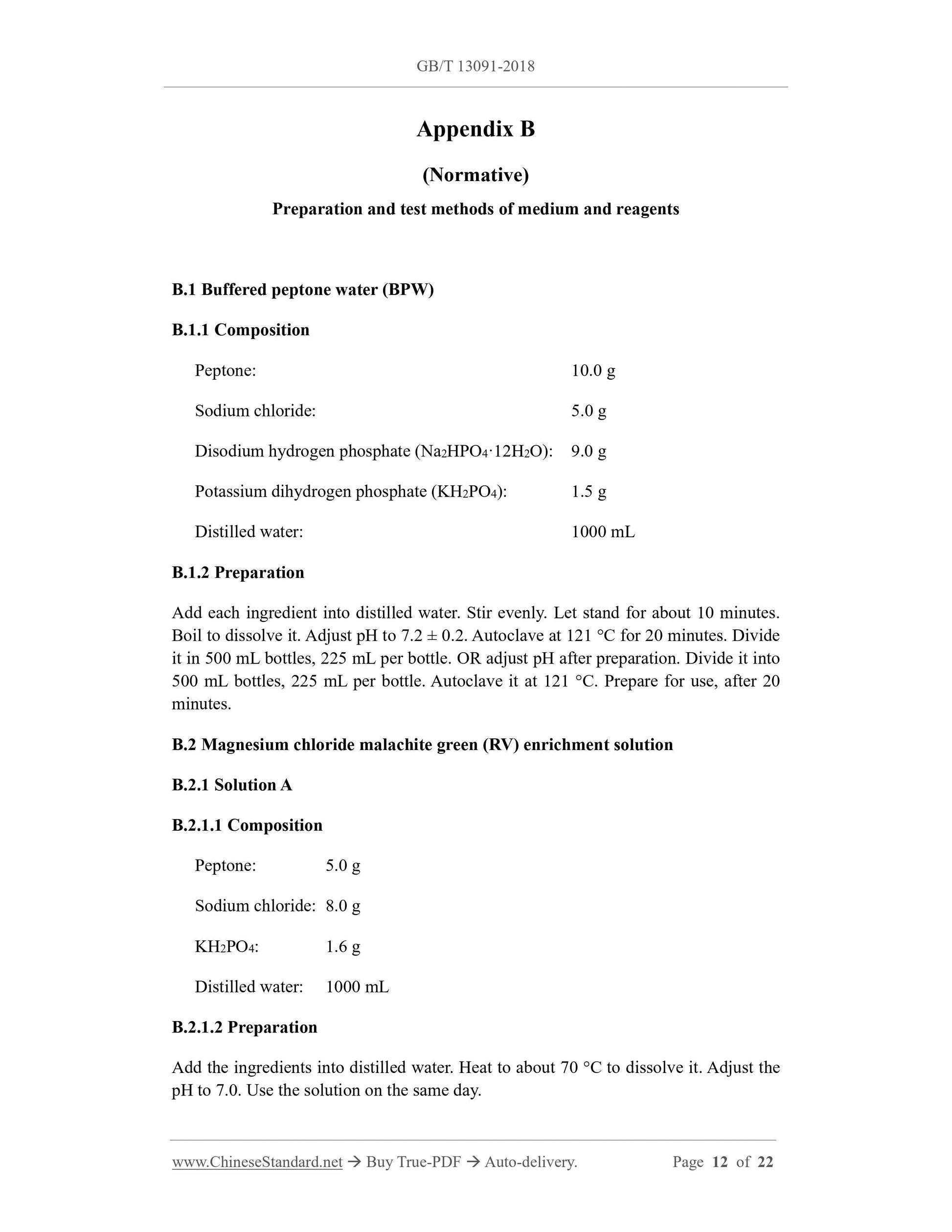
Appendix B
(Normative)
Preparation and test methods of medium and reagents
B.1 Buffered peptone water (BPW)
B.1.1 Composition
Peptone: 10.0 g
Sodium chloride: 5.0 g
Disodium hydrogen phosphate (Na2HPO4·12H2O): 9.0 g
Potassium dihydrogen phosphate (KH2PO4): 1.5 g
Distilled water: 1000 mL
B.1.2 Preparation
Add each ingredient into distilled water. Stir evenly. Let stand for about 10 minutes.
Boil to dissolve it. Adjust pH to 7.2 ± 0.2. Autoclave at 121 °C for 20 minutes. Divide
it in 500 mL bottles, 225 mL per bottle. OR adjust pH after preparation. Divide it into
500 mL bottles, 225 mL per bottle. Autoclave it at 121 °C. Prepare for use, after 20
minutes.
B.2 Magnesium chloride malachite green (RV) enrichment solution
B.2.1 Solution A
B.2.1.1 Composition
Peptone: 5.0 g
Sodium chloride: 8.0 g
KH2PO4: 1.6 g
Distilled water: 1000 mL
B.2.1.2 Preparation
Add the ingredients into distilled water. Heat to about 70 °C to dissolve it. Adjust the
pH to 7.0. Use the solution on the same day.
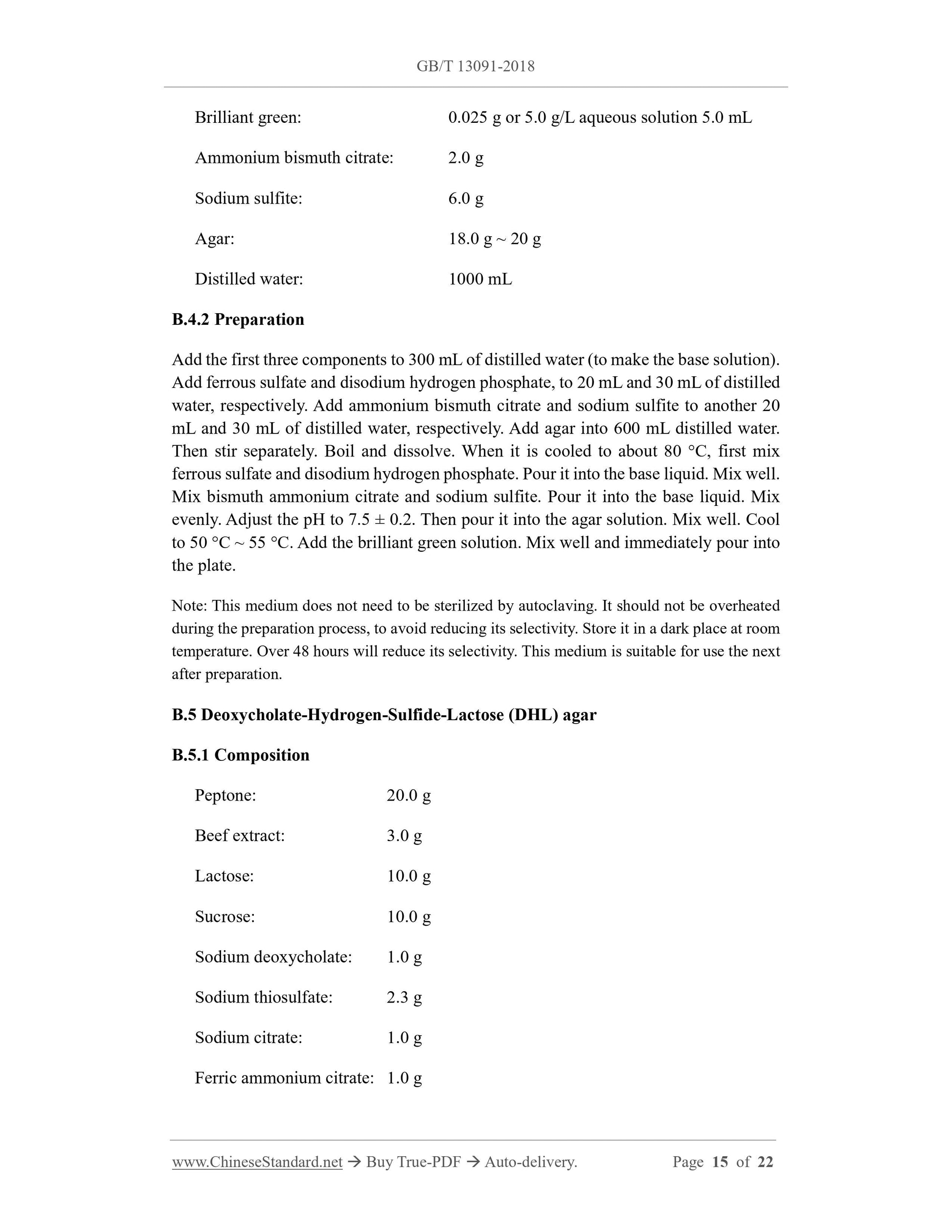
Brilliant green: 0.025 g or 5.0 g/L aqueous solution 5.0 mL
Ammonium bismuth citrate: 2.0 g
Sodium sulfite: 6.0 g
Agar: 18.0 g ~ 20 g
Distilled water: 1000 mL
B.4.2 Preparation
Add the first three components to 300 mL of distilled water (to make the base solution).
Add ferrous sulfate and disodium hydrogen phosphate, to 20 mL and 30 mL of distilled
water, respectively. Add ammonium bismuth citrate and sodium sulfite to another 20
mL and 30 mL of distilled water, respectively. Add agar into 600 mL distilled water.
Then stir separately. Boil and dissolve. When it is cooled to about 80 °C, first mix
ferrous sulfate and disodium hydrogen phosphate. Pour it into the base liquid. Mix well.
Mix bismuth ammonium citrate and sodium sulfite. Pour it into the base liquid. Mix
evenly. Adjust the pH to 7.5 ± 0.2. Then pour it into the agar solution. Mix well. Cool
to 50 °C ~ 55 °C. Add the brilliant green solution. Mix well and immediately pour into
the plate.
Note: This medium does not need to be sterilized by autoclaving. It should not be overheated
during the preparation process, to avoid reducing its selectivity. Store it in a dark place at room
temperature. Over 48 hours will reduce its selectivity. This medium is suitable for use the next
after preparation.
B.5 Deoxycholate-Hydrogen-Sulfide-Lactose (DHL) agar
B.5.1 Composition
Peptone: 20.0 g
Beef extract: 3.0 g
Lactose: 10.0 g
Sucrose: 10.0 g
Sodium deoxycholate: 1.0 g
Sodium thiosulfate: 2.3 g
Sodium citrate: 1.0 g
Ferric ammonium citrate: 1.0 g
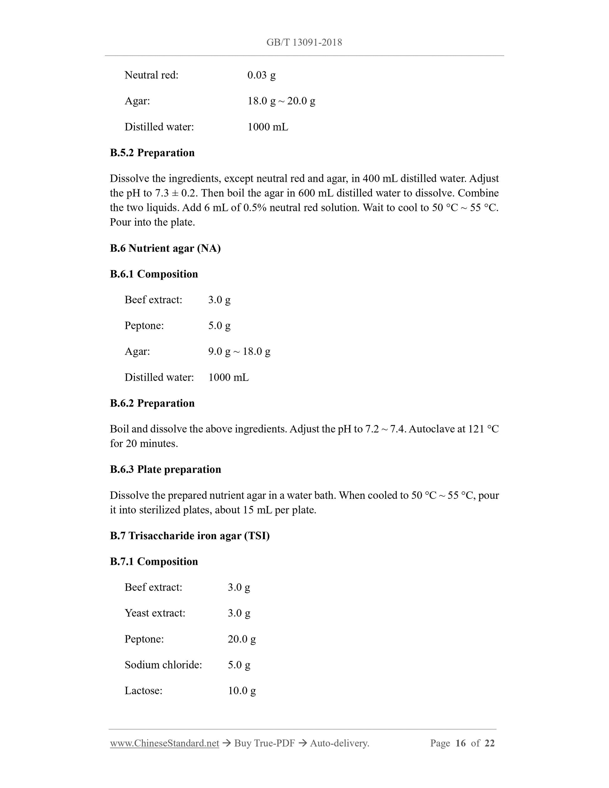
Neutral red: 0.03 g
Agar: 18.0 g ~ 20.0 g
Distilled water: 1000 mL
B.5.2 Preparation
Dissolve the ingredients, except neutral red and agar, in 400 mL distilled water. Adjust
the pH to 7.3 ± 0.2. Then boil the agar in 600 mL distilled water to dissolve. Combine
the two liquids. Add 6 mL of 0.5% neutral red solution. Wait to cool to 50 °C ~ 55 °C.
Pour into the plate.
B.6 Nutrient agar (NA)
B.6.1 Composition
Beef extract: 3.0 g
Peptone: 5.0 g
Agar: 9.0 g ~ 18.0 g
Distilled water: 1000 mL
B.6.2 Preparation
Boil and dissolve the above ingredients. Adjust the pH to 7.2 ~ 7.4. Autoclave at 121 °C
for 20 minutes.
B.6.3 Plate preparation
Dissolve the prepared nutrient agar in a water bath. When cooled to 50 °C ~ 55 °C, pour
it into sterilized plates, about 15 mL per plate.
B.7 Trisaccharide iron agar (TSI)
B.7.1 Composition
Beef extract: 3.0 g
Yeast extract: 3.0 g
Peptone: 20.0 g
Sodium chloride: 5.0 g
Lactose: 10.0 g
dissolved and cooled to 45 °C in advance. Divide it into test tubes. Place it on an
inclined plane for later use.
B.9.4 Test method
Pick the agar culture to inoculate. Incubate it at 36 °C ± 1 °C for 24 h. Observe the
results. Those with positive urease turns the culture medium red, due to alkali
production.
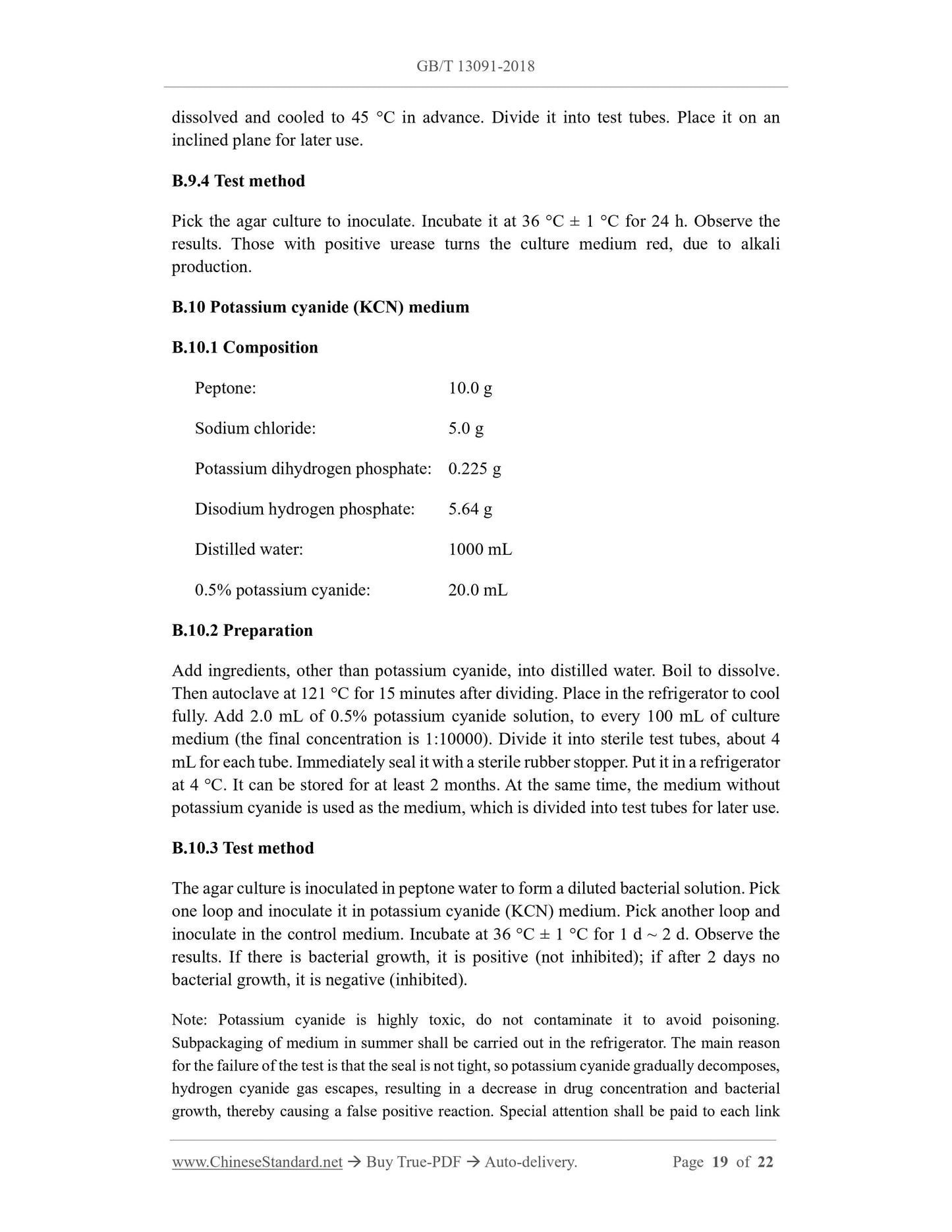
B.10 Potassium cyanide (KCN) medium
B.10.1 Composition
Peptone: 10.0 g
Sodium chloride: 5.0 g
Potassium dihydrogen phosphate: 0.225 g
Disodium hydrogen phosphate: 5.64 g
Distilled water: 1000 mL
0.5% potassium cyanide: 20.0 mL
B.10.2 Preparation
Add ingredients, other than potassium cyanide, into distilled water. Boil to dissolve.
Then autoclave at 121 °C for 15 minutes after dividing. Place in the refrigerator to cool
fully. Add 2.0 mL of 0.5% potassium cyanide solution, to every 100 mL of culture
medium (the final concentration is 1:10000). Divide it into sterile test tubes, about 4
mL for each tube. Immediately seal it with a sterile rubber stopper. Put it in a refrigerator
at 4 °C. It can be stored for at least 2 months. At the same time, the medium without
potassium cyanide is used as the medium, which is divided into test tubes for later use.
B.10.3 Test method
The agar culture is inoculated in peptone water to form a diluted bacterial solution. Pick
one loop and inoculate it in potassium cyanide (KCN) medium. Pick another loop and
inoculate in the control medium. Incubate at 36 °C ± 1 °C for 1 d ~ 2 d. Observe the
results. If there is bac...
Delivery: 9 seconds. Download (& Email) true-PDF + Invoice.
Get Quotation: Click GB/T 13091-2018 (Self-service in 1-minute)
Historical versions (Master-website): GB/T 13091-2018
Preview True-PDF (Reload/Scroll-down if blank)
GB/T 13091-2018
GB
NATIONAL STANDARD OF THE
PEOPLE’S REPUBLIC OF CHINA
ICS 65.120
B 46
Replacing GB/T 13091-2002
Determination of Salmonella in feeds
ISSUED ON: SEPTEMBER 17, 2018
IMPLEMENTED ON: APRIL 01, 2019
Issued by: State Administration for Market Regulation;
Standardization Administration of PRC.
Table of Contents
Foreword ... 3
1 Scope ... 4
2 Normative references ... 4
3 Medium or material... 4
4 Instruments and equipment ... 5
5 Samples ... 5
6 Test method (see Appendix A for the test procedure diagram) ... 6
7 Expression of results ... 10
Appendix A (Normative) Test procedure diagram ... 11
Appendix B (Normative) Preparation and test methods of medium and reagents ... 12
Determination of Salmonella in feeds
1 Scope
This standard specifies the inspection methods for Salmonella in feed.
This standard applies to the inspection of Salmonella in feed and feed additives.
2 Normative references
The following documents are essential to the application of this document. For the dated
documents, only the versions with the dates indicated are applicable to this document;
for the undated documents, only the latest version (including all the amendments) is
applicable to this standard.
GB/T 6682 Water for analytical laboratory use - Specification and test methods
3 Medium or material
Unless otherwise stated, only reagents confirmed to be of analytical grade are used in
the analysis.
3.1 Water: It shall meet the requirements of grade-3 water in GB/T 6682.
3.2 Buffered peptone water (BPW): See B.1 in Appendix B.
3.3 Magnesium chloride malachite green (RV) enrichment solution: See B.2 in
Appendix B.
3.4 Cystine selenite (SC) enrichment solution: See B.3 in Appendix B.
3.5 Bismuth sulfite (BS) agar: See B.4 in Appendix B.
3.6 Deoxycholate-Hydrogen-Sulfide-Lactose (DHL) agar: See B.5 in Appendix B.
3.7 Salmonella chromogenic medium.
3.8 Nutrient agar (NA): See B.6 in Appendix B.
3.9 Trisaccharide iron agar (TSI): See B.7 in Appendix B.
3.10 Peptone water and indigo-based reagents: See B.8 in Appendix B.
3.11 Urea agar (pH7.2): See B.9 in Appendix B.
3.12 Potassium cyanide (KCN) medium: See B.10 in Appendix B.
3.13 Lysine decarboxylase test medium: See B.11 in Appendix B.
3.14 Sugar fermentation tube: See B.12 in Appendix B.
3.15 Ortho-nitrophenol β-D-galactopyranoside (ONPG) medium: See B.13 in
Appendix B.
3.16 Semi-solid agar: See B.14 in Appendix B.
3.17 Sodium malonate medium: See B.15 in Appendix B.
3.18 Salmonella factor O and H polyvalent diagnostic serum, Vi factor diagnostic serum.
4 Instruments and equipment
4.1 Refrigerator: 2 °C ~ 5 °C.
4.2 Constant temperature incubator: 36 °C ± 1 °C, 42 °C ± 1 °C.
4.3 Homogenizer.
4.4 Oscillators.
4.5 Electronic balance: Sensitivity 0.1 g.
4.6 Sterile conical flasks: Capacity 500 mL, 250 mL, or sterile homogeneous bag.
4.7 Sterile pipettes: 1 mL (with 0.01 mL scale), 10 mL (with 0.1 mL scale), or
micropipette and tip.
4.8 Sterile petri dishes: Diameter 60 mm, 90 mm.
4.9 Sterile test tubes: 3 mm × 50 mm, 10 mm × 75 mm.
4.10 pH meter or pH colorimetric tube or precision pH test paper.
4.11 Automatic microbial biochemical identification system.
5 Samples
5.1 Sampling principles
The collection of samples shall follow the principles of randomness and
representativeness. The sampling process shall follow aseptic procedures, to prevent all
possible external contamination.
5.2 Sampling method
5.2.1 Samples shall be collected from the same batch of products. The sampling volume
of each sample shall meet the requirements for microbiological index inspection, which
is generally not less than 500 g (mL).
5.2.2 For solid products not larger than 500 g or liquid products not larger than 500 mL
in independent packaging, take the complete package.
5.2.3 For individually packaged liquid products larger than 500 mL, it shall be shaken
or stirred with a sterile stick before sampling, to make them uniform, then collect an
appropriate amount of samples and put them into a sterile sampling container, as a
sample.
5.2.4 For solid products with individual packages greater than 500 g, use a sterile
sampler to take appropriate samples from different parts of the same package; put them
into the same sterile sampling container, as one sample.
5.3 Storage and transport of collected samples
5.3.1 The samples shall be sent to the laboratory for testing, as soon as possible.
5.3.2 The samples shall be kept intact during transportation.
5.3.3 The samples shall be stored under conditions close to the original storage
temperature, OR necessary measures shall be taken to prevent changes in the number
of microorganisms in the samples.
6 Test method (see Appendix A for the test procedure
diagram)
6.1 Pre-enrichment
Weigh 25g (mL) of sample under sterile conditions. Add it into a 500 mL sterile conical
flask, which was filled with 225 mL of sterile BPW. Place it in an oscillator. Oscillate
at 8000 r/min ~ 10000 r/min for 2 min ~ 3 min. If the sample is in liquid form, shake
and mix well. Incubate it at 36 °C ± 1 °C for 18 h ± 2 h.
Note: A sterile homogenizing bag can be used instead of the conical flask. Then beating with a
homogenizer for 2 min ~ 3 min, but the homogenizing bag cannot be used for hard or angular
samples, only the conical flask can be used, to prevent the homogeneous bag from being
ruptured and leaked, in the homogenization and enrichment process, which may cause pollution.
b) Reaction No. A2: Supplementarily conduct mannitol and sorbitol tests; the results
of the two tests for the indigase-positive variant of Salmonella are all positive,
but it needs to be judged in combination with the results of serological
identification.
c) Reaction No. A3: Supplementarily conduct ONPG test. If ONPG is negative, then
it is Salmonella, while lysine decarboxylase is positive; if lysine decarboxylase is
negative, then it is Salmonella paratyphi A.
d) If necessary, carry out identification of Salmonella biochemical groups according
to Table 6.
6.4.3 If a biochemical identification kit or an automatic microbial biochemical
identification system is selected, suspicious colonies can be picked from the nutrient
agar plate, according to the preliminary judgment results in 6.4.1. A bacterial
suspension with appropriate turbidity can be prepared with physiological saline. Use a
biochemical identification kit or an automatic microbial biochemical identification
system for identification.
6.5 Serological identification
6.5.1 Checking the culture for autoagglutination
Use 1.2% ~ 1.5% agar culture as the antigen, for slide agglutination test. Firstly, to rule
out self-agglutination reaction, add a drop of normal saline to a clean glass slide; mix
the culture to be tested in the normal saline, to make a homogeneous turbid suspension;
shake the slide gently for 30 s ~ 60 s; observe the reaction in dark (observe with a
magnifying glass if necessary). If there is visible bacterial agglutination, it is considered
to have self-agglutination; otherwise, there is no self-agglutination. For the non-self-
agglutinating culture, refer to the following method for serological identification.
6.5.2 Identification of the O antigen
Draw 2 areas of about 1 cm × 2 cm on the slide. Pick 1 loop of bacteria to be tested. Put
1/2 loop on the upper part of each area on the slide. Add 1 drop of multivalent bacteria
O serum, to the lower part of one of the areas. Add 1 drop of normal saline to the lower
part of another area, as a control. Then use a sterile inoculation loop or needle, to grind
the colonies in the two areas into emulsion, respectively. Shake the slide for 1 min and
observe against the dark background. Any degree of agglutination is a positive reaction.
When the O serum does not agglutinate, inoculate the strain on a bacterial culture
medium, which has a high amount of agar (2% ~ 3%), to check again; if the O
agglutination reaction is prevented due to the presence of Vi antigen, the bacterial lawn
can be picked and placed in 1 mL normal saline, to make a concentrated bacterial
solution. Boil it on the flame of an alcohol lamp. Then check it.
6.5.3 Identification of H antigen
The operation is the same as 6.5.2. When the H antigen is underdeveloped, inoculate
the strain in the center of a 0.55% ~ 0.65% semi-solid agar plate. When the colony
spreads and grows, take bacteria from the edge for inspection. OR inoculate the strain
1 ~ 2 times in small glass tubes, which contain 0.3% ~ 0.4% semi-solid agar plate. Take
the bacteria from the far end for culture and re-inspection.
6.5.4 Identification of Vi antigen
The operation is the same as 6.5.2. Check with Vi factor serum.
7 Expression of results
Based on the results of the above biochemical tests and serological tests, it is concluded
that Salmonella is detected or not detected in 25g (mL) samples.
Appendix B
(Normative)
Preparation and test methods of medium and reagents
B.1 Buffered peptone water (BPW)
B.1.1 Composition
Peptone: 10.0 g
Sodium chloride: 5.0 g
Disodium hydrogen phosphate (Na2HPO4·12H2O): 9.0 g
Potassium dihydrogen phosphate (KH2PO4): 1.5 g
Distilled water: 1000 mL
B.1.2 Preparation
Add each ingredient into distilled water. Stir evenly. Let stand for about 10 minutes.
Boil to dissolve it. Adjust pH to 7.2 ± 0.2. Autoclave at 121 °C for 20 minutes. Divide
it in 500 mL bottles, 225 mL per bottle. OR adjust pH after preparation. Divide it into
500 mL bottles, 225 mL per bottle. Autoclave it at 121 °C. Prepare for use, after 20
minutes.
B.2 Magnesium chloride malachite green (RV) enrichment solution
B.2.1 Solution A
B.2.1.1 Composition
Peptone: 5.0 g
Sodium chloride: 8.0 g
KH2PO4: 1.6 g
Distilled water: 1000 mL
B.2.1.2 Preparation
Add the ingredients into distilled water. Heat to about 70 °C to dissolve it. Adjust the
pH to 7.0. Use the solution on the same day.
Brilliant green: 0.025 g or 5.0 g/L aqueous solution 5.0 mL
Ammonium bismuth citrate: 2.0 g
Sodium sulfite: 6.0 g
Agar: 18.0 g ~ 20 g
Distilled water: 1000 mL
B.4.2 Preparation
Add the first three components to 300 mL of distilled water (to make the base solution).
Add ferrous sulfate and disodium hydrogen phosphate, to 20 mL and 30 mL of distilled
water, respectively. Add ammonium bismuth citrate and sodium sulfite to another 20
mL and 30 mL of distilled water, respectively. Add agar into 600 mL distilled water.
Then stir separately. Boil and dissolve. When it is cooled to about 80 °C, first mix
ferrous sulfate and disodium hydrogen phosphate. Pour it into the base liquid. Mix well.
Mix bismuth ammonium citrate and sodium sulfite. Pour it into the base liquid. Mix
evenly. Adjust the pH to 7.5 ± 0.2. Then pour it into the agar solution. Mix well. Cool
to 50 °C ~ 55 °C. Add the brilliant green solution. Mix well and immediately pour into
the plate.
Note: This medium does not need to be sterilized by autoclaving. It should not be overheated
during the preparation process, to avoid reducing its selectivity. Store it in a dark place at room
temperature. Over 48 hours will reduce its selectivity. This medium is suitable for use the next
after preparation.
B.5 Deoxycholate-Hydrogen-Sulfide-Lactose (DHL) agar
B.5.1 Composition
Peptone: 20.0 g
Beef extract: 3.0 g
Lactose: 10.0 g
Sucrose: 10.0 g
Sodium deoxycholate: 1.0 g
Sodium thiosulfate: 2.3 g
Sodium citrate: 1.0 g
Ferric ammonium citrate: 1.0 g
Neutral red: 0.03 g
Agar: 18.0 g ~ 20.0 g
Distilled water: 1000 mL
B.5.2 Preparation
Dissolve the ingredients, except neutral red and agar, in 400 mL distilled water. Adjust
the pH to 7.3 ± 0.2. Then boil the agar in 600 mL distilled water to dissolve. Combine
the two liquids. Add 6 mL of 0.5% neutral red solution. Wait to cool to 50 °C ~ 55 °C.
Pour into the plate.
B.6 Nutrient agar (NA)
B.6.1 Composition
Beef extract: 3.0 g
Peptone: 5.0 g
Agar: 9.0 g ~ 18.0 g
Distilled water: 1000 mL
B.6.2 Preparation
Boil and dissolve the above ingredients. Adjust the pH to 7.2 ~ 7.4. Autoclave at 121 °C
for 20 minutes.
B.6.3 Plate preparation
Dissolve the prepared nutrient agar in a water bath. When cooled to 50 °C ~ 55 °C, pour
it into sterilized plates, about 15 mL per plate.
B.7 Trisaccharide iron agar (TSI)
B.7.1 Composition
Beef extract: 3.0 g
Yeast extract: 3.0 g
Peptone: 20.0 g
Sodium chloride: 5.0 g
Lactose: 10.0 g
dissolved and cooled to 45 °C in advance. Divide it into test tubes. Place it on an
inclined plane for later use.
B.9.4 Test method
Pick the agar culture to inoculate. Incubate it at 36 °C ± 1 °C for 24 h. Observe the
results. Those with positive urease turns the culture medium red, due to alkali
production.
B.10 Potassium cyanide (KCN) medium
B.10.1 Composition
Peptone: 10.0 g
Sodium chloride: 5.0 g
Potassium dihydrogen phosphate: 0.225 g
Disodium hydrogen phosphate: 5.64 g
Distilled water: 1000 mL
0.5% potassium cyanide: 20.0 mL
B.10.2 Preparation
Add ingredients, other than potassium cyanide, into distilled water. Boil to dissolve.
Then autoclave at 121 °C for 15 minutes after dividing. Place in the refrigerator to cool
fully. Add 2.0 mL of 0.5% potassium cyanide solution, to every 100 mL of culture
medium (the final concentration is 1:10000). Divide it into sterile test tubes, about 4
mL for each tube. Immediately seal it with a sterile rubber stopper. Put it in a refrigerator
at 4 °C. It can be stored for at least 2 months. At the same time, the medium without
potassium cyanide is used as the medium, which is divided into test tubes for later use.
B.10.3 Test method
The agar culture is inoculated in peptone water to form a diluted bacterial solution. Pick
one loop and inoculate it in potassium cyanide (KCN) medium. Pick another loop and
inoculate in the control medium. Incubate at 36 °C ± 1 °C for 1 d ~ 2 d. Observe the
results. If there is bac...
Share