


1
/
of
12
www.ChineseStandard.us -- Field Test Asia Pte. Ltd.
YY/T 0744-2018 English PDF (YY/T0744-2018)
YY/T 0744-2018 English PDF (YY/T0744-2018)
Regular price
$380.00
Regular price
Sale price
$380.00
Unit price
/
per
Shipping calculated at checkout.
Couldn't load pickup availability
YY/T 0744-2018: Particular specifications for mobile C-arm X-ray equipment
Delivery: 9 seconds. Download (& Email) true-PDF + Invoice.
Get Quotation: Click YY/T 0744-2018 (Self-service in 1-minute)
Historical versions (Master-website): YY/T 0744-2018
Preview True-PDF (Reload/Scroll-down if blank)
YY/T 0744-2018
PHARMACEUTICAL INDUSTRY STANDARD
OF THE PEOPLE’S REPUBLIC OF CHINA
ICS 11.040.50
C 43
Replacing YY/T 0744-2009
Particular specifications for mobile C-arm X-ray equipment
ISSUED ON: JANUARY 19, 2018
IMPLEMENTED ON: JANUARY 01, 2019
Issued by: China Food and Drug Administration
Table of Contents
Foreword ... 3
1 Scope ... 5
2 Normative references ... 5
3 Terms and definitions... 6
4 Classification and composition ... 7
5 Requirements ... 8
6 Test methods ... 19
Appendix A (Normative) Test layout ... 31
Appendix B (Normative) Test phantom ... 34
Appendix C (Normative) Test method of correction factor g ... 39
Particular specifications for mobile C-arm X-ray equipment
1 Scope
This standard specifies the terms and definitions, classification and composition,
requirements and test methods of mobile C-arm X-ray equipments (hereinafter referred
to as C-arm X-ray equipments).
This standard applies to mobile X-ray equipments with C-arm mechanical support
devices, which are mainly used for positioning and inspection, in surgical operations in
medical and health institutions. This standard does not apply to C-arm X-ray equipment,
which has a maximum focus-image-receiver distance (SID) of less than 60 cm.
2 Normative references
The following documents are essential to the application of this document. For the dated
documents, only the versions with the dates indicated are applicable to this document;
for the undated documents, only the latest version (including all the amendments) is
applicable to this standard.
GB 9706.1-2007 Medical electrical equipment - Part 1: General requirements for
safety
GB 9706.3-2000 Medical electrical equipment - Part 2: Particular requirements for
the safety of high-voltage generators of diagnostic X-ray generators
GB 9706.11-1997 Medical electrical equipment - Part 2: Particular requirements for
the safety of X-ray source assemblies and X-ray tube assemblies for medical
diagnosis
GB 9706.12-1997 Medical electrical equipment - Part 1: General requirements for
safety - 3. Collateral standard: General requirements for radiation protection in
diagnostic X-ray equipment
GB 9706.14-1997 Medical electrical equipment - Part 2: Particular requirements for
the safety of associated equipment of X-ray equipment
GB 9706.15-2008 Medical electrical equipment - Part 1: General requirements for
safety - 1. Collateral standard: Safety requirements for medical electrical systems
GB 9706.23-2005 Medical electrical equipment - Part 2-43: Particular requirements
for the safety of X-ray equipment for interventional procedures
GB/T 10149 Terminology and symbol for medical X-ray equipment
GB/T 10151 Medical diagnostic X-ray equipment - Specifications for high voltage
cable plugs and sockets
YY 0076-1992 Coating classifications for metal product - Technical conditions
YY/T 0106-2008 General specifications for medical diagnostic X-ray equipment
YY/T 0291 Environmental requirements and test methods for medical X-ray
equipment
YY 0505 Medical electrical equipment - Part 1-2: General requirements for safety -
Collateral standards: Electromagnetic compatibility - Requirements and tests
IEC 61910-1:2014 Medical electrical equipment - Radiation dose documentation -
Part 1: Radiation dose structured reports for radiography and radioscopy
3 Terms and definitions
The terms and definitions, which are defined in GB/T 10149, as well as the following
terms and definitions, apply to this document.
3.1
Fluoroscopy spot image
An image, which is obtained by superimposing multiple frames of fluoroscopic
images collected (has a higher signal-to-noise ratio than ordinary fluoroscopic
images).
3.2
Fluoroscopy image subtraction
A digital image processing method, for subtracting fluoroscopic sequence images.
3.3
Radiography image subtraction
Digital image processing method, for subtraction of radiographic sequence images.
Note 1: Compared with "fluoroscopy image subtraction", "radiography image subtraction"
has a higher X-ray dose and a higher quality of the subtracted image.
5 Requirements
5.1 Working conditions
5.1.1 Environmental conditions
Unless otherwise specified, the working environmental conditions of the C-arm X-ray
equipment shall meet the following requirements:
a) Ambient temperature: 10 °C ~ 40 °C;
b) Relative humidity: 30% ~ 75%;
c) Atmospheric pressure: 700 hPa ~ 1060 hPa.
5.1.2 Power conditions
The manufacturer shall describe the power supply conditions, which are used by the
product, in the accompanying documents. The working power supply conditions shall
meet:
a) Power supply voltage and number of phases: As specified by the manufacturer,
the network voltage fluctuation shall not exceed ±10% of the nominal value;
b) Power frequency: 50 Hz ± 1 Hz;
c) Power supply resistance: As specified by the manufacturer, but shall meet the
requirements of Table 101 in 10.2.2a) of GB 9706.3-2000;
d) Power capacity: As specified by the manufacturer.
5.2 Electric power
5.2.1 Maximum output electrical power
A C-arm X-ray equipment shall specify the corresponding combination of X-ray tube
voltage and X-ray tube current, that results in maximum output electrical power, in
fluoroscopy and/or radiography modes.
5.2.2 Nominal electrical power
It shall be stipulated that when the loading time is 0.1 s and the X-ray tube voltage is
100 kV, the maximum constant electric power output, expressed in kilowatts (kW), that
can be provided by the X-ray generating device, as the nominal electric power. If this
value cannot be pre-selected, it may use the X-ray tube voltage value, which is closest
to 100 kV and the closest loading time value, but not shorter than 0.1 s.
The nominal electrical power shall be given, together with a combination of X-ray tube
voltage, X-ray tube current, loading time.
5.3 Loading factors and controls
5.3.1 X-ray tube voltage
The X-ray tube voltage shall meet the following requirements:
a) The manufacturer shall specify the adjustment range and adjustment method of
the X-ray tube voltage;
b) The manufacturer shall specify the deviation of the X-ray tube voltage value,
which shall at least meet the requirements of 50.103.1 in GB 9706.3-2000.
5.3.2 X-ray tube current
The X-ray tube current shall meet the following requirements:
a) The manufacturer shall specify the adjustment range and adjustment method of
X-ray tube current, in continuous and highest frame rate pulsed fluoroscopy mode;
b) The manufacturer shall specify the deviation of the current value of the X-ray tube,
which shall at least meet the requirements of 50.103.2 in GB 9706.3-2000.
5.3.3 Loading time
The loading time shall meet the following requirements:
a) If the loading time display is provided and the single-frame radiography function
is provided, the manufacturer shall specify the adjustment range and adjustment
method of the loading time, for single-frame radiography;
b) The manufacturer shall specify the deviation of the loading time value, which
shall at least meet the requirements of 50.103.3 in GB 9706.3-2000.
5.3.4 Current time product
If the current time product display is provided, the current time product shall meet the
following requirements:
a) The manufacturer shall specify the adjustment range and adjustment method of
current time product;
b) The manufacturer shall specify the deviation of the current time product, which
shall at least meet the requirements of 50.103.4 in GB 9706.3-2000.
5.3.5 Overload protection
shall not be greater than 5 s.
5.4.8 Fluoroscopy imaging stability time
The manufacturer shall specify the fluoroscopic image stabilization time, under
continuous and highest pulse fluoroscopic frame rate, which shall not exceed 2 s.
5.4.9 Fluoroscopy recovery time
The fluoroscopy recovery time shall not exceed 5 min.
5.4.10 Fluoroscopic performance during network transmission
In fluoroscopy, if selecting network transmission, it shall not affect the fluoroscopy.
5.4.11 Artifacts
No artifacts affecting clinical application.
5.4.12 Digital subtraction imaging performance
5.4.12.1 Imaging performance of radiography image subtraction
If the C-arm X-ray equipment has the radiography image subtraction imaging function,
it shall meet the following requirements:
a) The manufacturer shall specify the dynamic range; but at least the thickest one
vessel simulation component is visible in all steps;
b) The manufacturer shall specify the contrast sensitivity; but the 0.1 mm vessel
simulation component shall be visible, at least in the 0.8 mm copper step;
c) When the two images of the fixed object have the same spatial coordinate
characteristics, there shall be no misregistration artifacts.
5.4.12.2 Imaging performance of fluoroscopy image subtraction
If the C-arm X-ray equipment has the function of fluoroscopic image subtraction
imaging, it shall meet the following requirements:
a) The manufacturer shall specify the contrast sensitivity; but the 0.4 mm vessel
simulation component shall be visible, at least in the 0.8 mm copper step.
b) When the two images of the fixed object have the same spatial coordinate
characteristics, there shall be no misregistration artifacts.
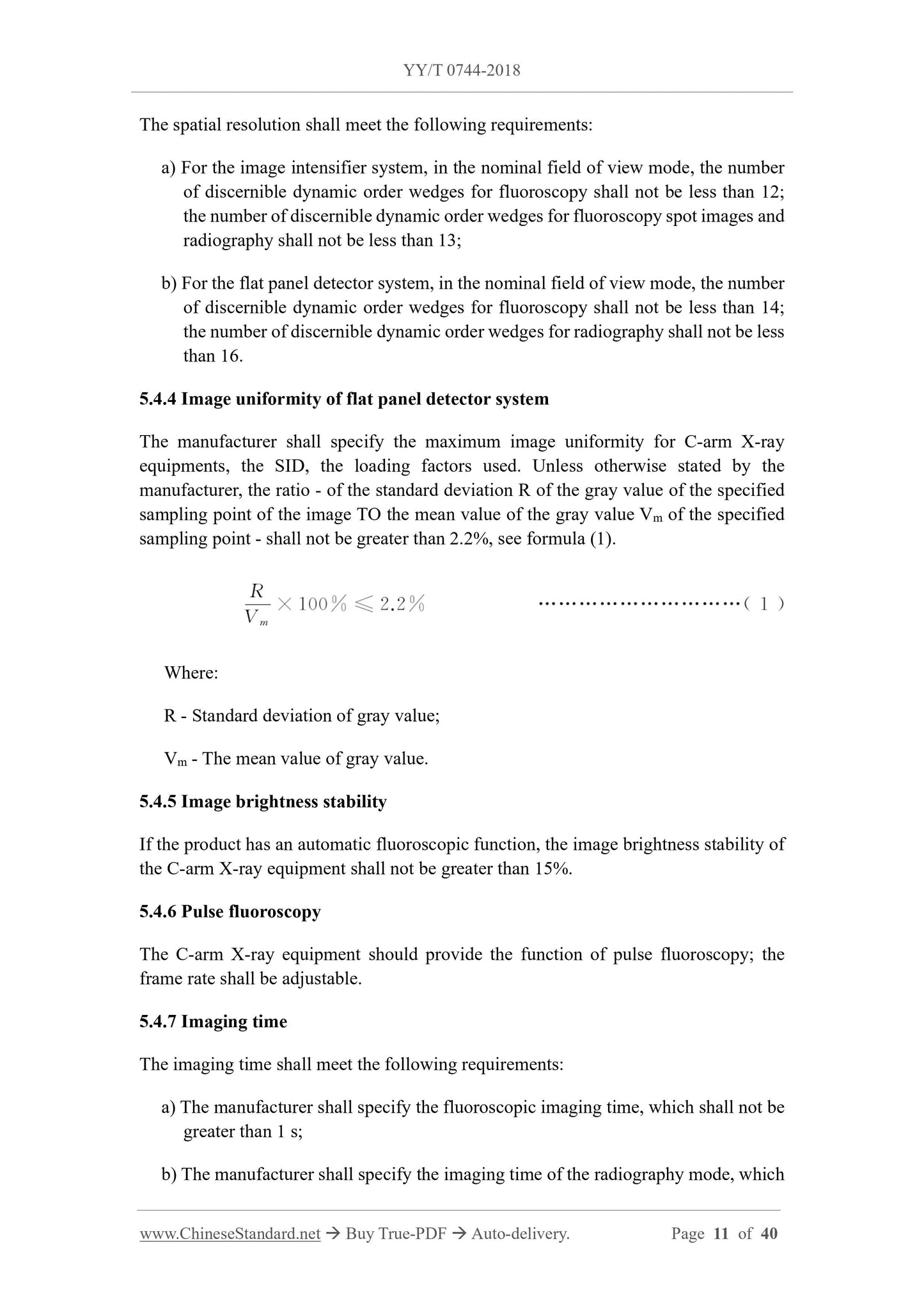
5.4.13 3D imaging
5.4.13.1 3D acquisition parameters
The manufacturer shall specify the three-dimensional acquisition angle, the number of
acquired image frames, the size of the reconstruction area, where the acquisition angle
should not be less than 180°.
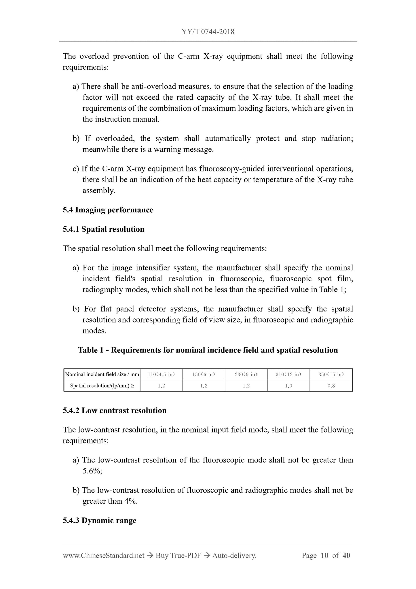
5.4.13.2 3D imaging spatial resolution
The manufacturer shall specify the cross-sectional spatial resolution of the three-
dimensional imaging. If not specified, at least 4 independent holes of 1.3 mm shall be
seen, in the high-contrast mode, OR at least 4 independent holes of 0.9 mm shall be
seen, in the low-contrast mode.
Note: See 5.5.2 for dose requirements for high or low contrast mode of 3D imaging.
5.4.13.3 3D image artifacts
There shall be no artifacts, which affect the clinical application.
5.5 Radiation safety
5.5.1 Dosage indication
The dose indication of the C-arm X-ray equipment shall meet the following
requirements:
a) The instruction manual shall specify the measurement reference point and
calibration information of the dose indication value.
b) During the fluoroscopy process, the air kerma rate shall be continuously indicated
in milliGe per minute (mGy/min); refreshed at least once per second; the
indication shall be clearly visible in the normal working position.
c) Since the last reset setting, the cumulative value of air kerma, which is generated
by all fluoroscopy and radiography shall be continuously indicated in milliGe
(mGy); refreshed at least every 5s; OR indicated within 5 s after the end of loading,
meanwhile the indication shall be clearly visible in the normal working position.
d) The indicated values of air kerma rate and accumulated air kerma shall be clearly
distinguishable.
e) Accuracy of air kerma and air kerma rate:
- The error of the indicated value of air kerma rate, which exceeds 6 mGy/min,
shall not be greater than 35%;
- The error of the indicated value of accumulated air kerma energy, which exceeds
100 mGy, shall not be greater than 35%.
f) The C-arm X-ray equipment shall provide an indication of the cumulative dose
meet the requirements of radiation dose for medical X-ray radiography and fluoroscopy
equipment in IEC 61910-1:2014.
5.5.5 Pediatric examinations
If the equipment is claimed to be suitable for use as a pediatric examination, it shall
meet the following requirements:
a) Instructions for use shall include measures for dose reduction in pediatric
examinations;
b) The anti-scatter grid should be detachable without tools, BUT it is not suitable for
C-arm X-ray equipments, that can be used with navigation equipment.
5.6 Mechanical device performance
5.6.1 Mechanical range of movement
5.6.1.1 Sliding of C-arm
The equipment, whose C-arm can slide along the arc of the track, shall meet the
following requirements:
a) The arc sliding angle along the track should not be less than 110°. If there is an
angle indication, the deviation, between the indicated value and the actual value,
shall be specified by the manufacturer;
b) The manually operated C-arm sliding shall be provided with a locking and
positioning device; its braking torque shall not be less than 50 N·m;
c) Manually operated slipping start torque shall not be greater than 40 N·m.
5.6.1.2 C-arm rotation
Equipment, whose C-arm can be rotated along the horizontal axis, shall meet the
following requirements:
a) The rotation angle should not be less than ±180°. If there is an angle indication,
the deviation, between the indicated value and the actual value, shall be specified
by the manufacturer;
b) The manually operated C-arm rotation shall be provided with a locking and
positioning device; its braking torque shall not be less than 45 N·m;
c) The rotation starting torque of manual operation shall not be greater than 35 N·m.
5.6.1.3 Movement of C-arm
Equipment, whose C-arm can move vertically and horizontally, shall meet the following
requirements:
a) The movement range shall be specified; if there is an indication, the deviation of
the indicated value shall be specified by the manufacturer;
b) The movement of manually operated C-arm shall be provided with a locking and
positioning device; its braking force shall not be less than 100 N;
c) The starting force shall meet the requirements of 5.5.5 in YY/T 0106-2008.
5.6.1.4 Horizontal swing
For the equipment, whose C-arm can be used for horizontal swing, its swing shall meet
the following requirements:
a) The manufacturer shall specify the swing angle and the deviation value of the
angle;
b) The swing of manually operated C-arm shall be provided with a locking and
positioning device; its braking torque shall not be less than 40 N·m;
c) The swing starting torque of manual operation sha...
Delivery: 9 seconds. Download (& Email) true-PDF + Invoice.
Get Quotation: Click YY/T 0744-2018 (Self-service in 1-minute)
Historical versions (Master-website): YY/T 0744-2018
Preview True-PDF (Reload/Scroll-down if blank)
YY/T 0744-2018
PHARMACEUTICAL INDUSTRY STANDARD
OF THE PEOPLE’S REPUBLIC OF CHINA
ICS 11.040.50
C 43
Replacing YY/T 0744-2009
Particular specifications for mobile C-arm X-ray equipment
ISSUED ON: JANUARY 19, 2018
IMPLEMENTED ON: JANUARY 01, 2019
Issued by: China Food and Drug Administration
Table of Contents
Foreword ... 3
1 Scope ... 5
2 Normative references ... 5
3 Terms and definitions... 6
4 Classification and composition ... 7
5 Requirements ... 8
6 Test methods ... 19
Appendix A (Normative) Test layout ... 31
Appendix B (Normative) Test phantom ... 34
Appendix C (Normative) Test method of correction factor g ... 39
Particular specifications for mobile C-arm X-ray equipment
1 Scope
This standard specifies the terms and definitions, classification and composition,
requirements and test methods of mobile C-arm X-ray equipments (hereinafter referred
to as C-arm X-ray equipments).
This standard applies to mobile X-ray equipments with C-arm mechanical support
devices, which are mainly used for positioning and inspection, in surgical operations in
medical and health institutions. This standard does not apply to C-arm X-ray equipment,
which has a maximum focus-image-receiver distance (SID) of less than 60 cm.
2 Normative references
The following documents are essential to the application of this document. For the dated
documents, only the versions with the dates indicated are applicable to this document;
for the undated documents, only the latest version (including all the amendments) is
applicable to this standard.
GB 9706.1-2007 Medical electrical equipment - Part 1: General requirements for
safety
GB 9706.3-2000 Medical electrical equipment - Part 2: Particular requirements for
the safety of high-voltage generators of diagnostic X-ray generators
GB 9706.11-1997 Medical electrical equipment - Part 2: Particular requirements for
the safety of X-ray source assemblies and X-ray tube assemblies for medical
diagnosis
GB 9706.12-1997 Medical electrical equipment - Part 1: General requirements for
safety - 3. Collateral standard: General requirements for radiation protection in
diagnostic X-ray equipment
GB 9706.14-1997 Medical electrical equipment - Part 2: Particular requirements for
the safety of associated equipment of X-ray equipment
GB 9706.15-2008 Medical electrical equipment - Part 1: General requirements for
safety - 1. Collateral standard: Safety requirements for medical electrical systems
GB 9706.23-2005 Medical electrical equipment - Part 2-43: Particular requirements
for the safety of X-ray equipment for interventional procedures
GB/T 10149 Terminology and symbol for medical X-ray equipment
GB/T 10151 Medical diagnostic X-ray equipment - Specifications for high voltage
cable plugs and sockets
YY 0076-1992 Coating classifications for metal product - Technical conditions
YY/T 0106-2008 General specifications for medical diagnostic X-ray equipment
YY/T 0291 Environmental requirements and test methods for medical X-ray
equipment
YY 0505 Medical electrical equipment - Part 1-2: General requirements for safety -
Collateral standards: Electromagnetic compatibility - Requirements and tests
IEC 61910-1:2014 Medical electrical equipment - Radiation dose documentation -
Part 1: Radiation dose structured reports for radiography and radioscopy
3 Terms and definitions
The terms and definitions, which are defined in GB/T 10149, as well as the following
terms and definitions, apply to this document.
3.1
Fluoroscopy spot image
An image, which is obtained by superimposing multiple frames of fluoroscopic
images collected (has a higher signal-to-noise ratio than ordinary fluoroscopic
images).
3.2
Fluoroscopy image subtraction
A digital image processing method, for subtracting fluoroscopic sequence images.
3.3
Radiography image subtraction
Digital image processing method, for subtraction of radiographic sequence images.
Note 1: Compared with "fluoroscopy image subtraction", "radiography image subtraction"
has a higher X-ray dose and a higher quality of the subtracted image.
5 Requirements
5.1 Working conditions
5.1.1 Environmental conditions
Unless otherwise specified, the working environmental conditions of the C-arm X-ray
equipment shall meet the following requirements:
a) Ambient temperature: 10 °C ~ 40 °C;
b) Relative humidity: 30% ~ 75%;
c) Atmospheric pressure: 700 hPa ~ 1060 hPa.
5.1.2 Power conditions
The manufacturer shall describe the power supply conditions, which are used by the
product, in the accompanying documents. The working power supply conditions shall
meet:
a) Power supply voltage and number of phases: As specified by the manufacturer,
the network voltage fluctuation shall not exceed ±10% of the nominal value;
b) Power frequency: 50 Hz ± 1 Hz;
c) Power supply resistance: As specified by the manufacturer, but shall meet the
requirements of Table 101 in 10.2.2a) of GB 9706.3-2000;
d) Power capacity: As specified by the manufacturer.
5.2 Electric power
5.2.1 Maximum output electrical power
A C-arm X-ray equipment shall specify the corresponding combination of X-ray tube
voltage and X-ray tube current, that results in maximum output electrical power, in
fluoroscopy and/or radiography modes.
5.2.2 Nominal electrical power
It shall be stipulated that when the loading time is 0.1 s and the X-ray tube voltage is
100 kV, the maximum constant electric power output, expressed in kilowatts (kW), that
can be provided by the X-ray generating device, as the nominal electric power. If this
value cannot be pre-selected, it may use the X-ray tube voltage value, which is closest
to 100 kV and the closest loading time value, but not shorter than 0.1 s.
The nominal electrical power shall be given, together with a combination of X-ray tube
voltage, X-ray tube current, loading time.
5.3 Loading factors and controls
5.3.1 X-ray tube voltage
The X-ray tube voltage shall meet the following requirements:
a) The manufacturer shall specify the adjustment range and adjustment method of
the X-ray tube voltage;
b) The manufacturer shall specify the deviation of the X-ray tube voltage value,
which shall at least meet the requirements of 50.103.1 in GB 9706.3-2000.
5.3.2 X-ray tube current
The X-ray tube current shall meet the following requirements:
a) The manufacturer shall specify the adjustment range and adjustment method of
X-ray tube current, in continuous and highest frame rate pulsed fluoroscopy mode;
b) The manufacturer shall specify the deviation of the current value of the X-ray tube,
which shall at least meet the requirements of 50.103.2 in GB 9706.3-2000.
5.3.3 Loading time
The loading time shall meet the following requirements:
a) If the loading time display is provided and the single-frame radiography function
is provided, the manufacturer shall specify the adjustment range and adjustment
method of the loading time, for single-frame radiography;
b) The manufacturer shall specify the deviation of the loading time value, which
shall at least meet the requirements of 50.103.3 in GB 9706.3-2000.
5.3.4 Current time product
If the current time product display is provided, the current time product shall meet the
following requirements:
a) The manufacturer shall specify the adjustment range and adjustment method of
current time product;
b) The manufacturer shall specify the deviation of the current time product, which
shall at least meet the requirements of 50.103.4 in GB 9706.3-2000.
5.3.5 Overload protection
shall not be greater than 5 s.
5.4.8 Fluoroscopy imaging stability time
The manufacturer shall specify the fluoroscopic image stabilization time, under
continuous and highest pulse fluoroscopic frame rate, which shall not exceed 2 s.
5.4.9 Fluoroscopy recovery time
The fluoroscopy recovery time shall not exceed 5 min.
5.4.10 Fluoroscopic performance during network transmission
In fluoroscopy, if selecting network transmission, it shall not affect the fluoroscopy.
5.4.11 Artifacts
No artifacts affecting clinical application.
5.4.12 Digital subtraction imaging performance
5.4.12.1 Imaging performance of radiography image subtraction
If the C-arm X-ray equipment has the radiography image subtraction imaging function,
it shall meet the following requirements:
a) The manufacturer shall specify the dynamic range; but at least the thickest one
vessel simulation component is visible in all steps;
b) The manufacturer shall specify the contrast sensitivity; but the 0.1 mm vessel
simulation component shall be visible, at least in the 0.8 mm copper step;
c) When the two images of the fixed object have the same spatial coordinate
characteristics, there shall be no misregistration artifacts.
5.4.12.2 Imaging performance of fluoroscopy image subtraction
If the C-arm X-ray equipment has the function of fluoroscopic image subtraction
imaging, it shall meet the following requirements:
a) The manufacturer shall specify the contrast sensitivity; but the 0.4 mm vessel
simulation component shall be visible, at least in the 0.8 mm copper step.
b) When the two images of the fixed object have the same spatial coordinate
characteristics, there shall be no misregistration artifacts.
5.4.13 3D imaging
5.4.13.1 3D acquisition parameters
The manufacturer shall specify the three-dimensional acquisition angle, the number of
acquired image frames, the size of the reconstruction area, where the acquisition angle
should not be less than 180°.
5.4.13.2 3D imaging spatial resolution
The manufacturer shall specify the cross-sectional spatial resolution of the three-
dimensional imaging. If not specified, at least 4 independent holes of 1.3 mm shall be
seen, in the high-contrast mode, OR at least 4 independent holes of 0.9 mm shall be
seen, in the low-contrast mode.
Note: See 5.5.2 for dose requirements for high or low contrast mode of 3D imaging.
5.4.13.3 3D image artifacts
There shall be no artifacts, which affect the clinical application.
5.5 Radiation safety
5.5.1 Dosage indication
The dose indication of the C-arm X-ray equipment shall meet the following
requirements:
a) The instruction manual shall specify the measurement reference point and
calibration information of the dose indication value.
b) During the fluoroscopy process, the air kerma rate shall be continuously indicated
in milliGe per minute (mGy/min); refreshed at least once per second; the
indication shall be clearly visible in the normal working position.
c) Since the last reset setting, the cumulative value of air kerma, which is generated
by all fluoroscopy and radiography shall be continuously indicated in milliGe
(mGy); refreshed at least every 5s; OR indicated within 5 s after the end of loading,
meanwhile the indication shall be clearly visible in the normal working position.
d) The indicated values of air kerma rate and accumulated air kerma shall be clearly
distinguishable.
e) Accuracy of air kerma and air kerma rate:
- The error of the indicated value of air kerma rate, which exceeds 6 mGy/min,
shall not be greater than 35%;
- The error of the indicated value of accumulated air kerma energy, which exceeds
100 mGy, shall not be greater than 35%.
f) The C-arm X-ray equipment shall provide an indication of the cumulative dose
meet the requirements of radiation dose for medical X-ray radiography and fluoroscopy
equipment in IEC 61910-1:2014.
5.5.5 Pediatric examinations
If the equipment is claimed to be suitable for use as a pediatric examination, it shall
meet the following requirements:
a) Instructions for use shall include measures for dose reduction in pediatric
examinations;
b) The anti-scatter grid should be detachable without tools, BUT it is not suitable for
C-arm X-ray equipments, that can be used with navigation equipment.
5.6 Mechanical device performance
5.6.1 Mechanical range of movement
5.6.1.1 Sliding of C-arm
The equipment, whose C-arm can slide along the arc of the track, shall meet the
following requirements:
a) The arc sliding angle along the track should not be less than 110°. If there is an
angle indication, the deviation, between the indicated value and the actual value,
shall be specified by the manufacturer;
b) The manually operated C-arm sliding shall be provided with a locking and
positioning device; its braking torque shall not be less than 50 N·m;
c) Manually operated slipping start torque shall not be greater than 40 N·m.
5.6.1.2 C-arm rotation
Equipment, whose C-arm can be rotated along the horizontal axis, shall meet the
following requirements:
a) The rotation angle should not be less than ±180°. If there is an angle indication,
the deviation, between the indicated value and the actual value, shall be specified
by the manufacturer;
b) The manually operated C-arm rotation shall be provided with a locking and
positioning device; its braking torque shall not be less than 45 N·m;
c) The rotation starting torque of manual operation shall not be greater than 35 N·m.
5.6.1.3 Movement of C-arm
Equipment, whose C-arm can move vertically and horizontally, shall meet the following
requirements:
a) The movement range shall be specified; if there is an indication, the deviation of
the indicated value shall be specified by the manufacturer;
b) The movement of manually operated C-arm shall be provided with a locking and
positioning device; its braking force shall not be less than 100 N;
c) The starting force shall meet the requirements of 5.5.5 in YY/T 0106-2008.
5.6.1.4 Horizontal swing
For the equipment, whose C-arm can be used for horizontal swing, its swing shall meet
the following requirements:
a) The manufacturer shall specify the swing angle and the deviation value of the
angle;
b) The swing of manually operated C-arm shall be provided with a locking and
positioning device; its braking torque shall not be less than 40 N·m;
c) The swing starting torque of manual operation sha...
Share